did-you-know? rent-now

Amazon no longer offers textbook rentals. We do!

did-you-know? rent-now

Amazon no longer offers textbook rentals. We do!

We're the #1 textbook rental company. Let us show you why.

9780683301182

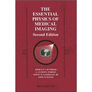
The Essential Physics of Medical Imaging

by ; ; ;
  • ISBN13:

    9780683301182

  • ISBN10:

    0683301187

  • Edition: 2nd
  • Format: Hardcover
  • Copyright: 2001-11-16
  • Publisher: LWW
  • View Upgraded Edition

Note: Supplemental materials are not guaranteed with Rental or Used book purchases.

Purchase Benefits

  • Free Shipping Icon Free Shipping On Orders Over $35!
    Your order must be $35 or more to qualify for free economy shipping. Bulk sales, PO's, Marketplace items, eBooks and apparel do not qualify for this offer.
  • eCampus.com Logo Get Rewarded for Ordering Your Textbooks! Enroll Now
List Price: $169.00 Save up to $42.25
  • Buy Used
    $126.75
    Add to Cart Free Shipping Icon Free Shipping

    USUALLY SHIPS IN 2-4 BUSINESS DAYS

Supplemental Materials

What is included with this book?

Summary

Developed from the authors' highly successful annual imaging physics review course, this new Second Edition gives readers a clear, fundamental understanding of the theory and applications of physics in radiology, nuclear medicine, and radiobiology. The Essential Physics of Medical Imaging, Second Edition provides key coverage of the clinical implications of technical principles--making this book great for board review. Highlights of this new edition include completely updated and expanded chapters and more than 960 illustrations. Major sections cover basic concepts, diagnostic radiology, nuclear medicine, and radiation protection, dosimetry, and biology. A Brandon-Hill recommended title.

Table of Contents

Preface xv
Acknowledgments xvii
Foreword xix
SECTION I: BASIC CONCEPTS 1(94)
Introduction to Medical Imaging
3(14)
The Modalities
4(9)
Image Properties
13(4)
Radiation and the Atom
17(14)
Radiation
17(4)
Structure of the Atom
21(10)
Interaction of Radiation with Matter
31(30)
Particle Interactions
31(6)
X- and Gamma Ray Interactions
37(8)
Attenuation of X- and Gamma Rays
45(7)
Absorption of Energy from X- and Gamma Rays
52(4)
Imparted Energy, Equivalent Dose, and Effective Dose
56(5)
Computers in Medical Imaging
61(34)
Storage and Transfer of Data in Computers
61(5)
Analog Data and Conversion between Analog and Digital Forms
66(4)
Components and Operation of Computers
70(8)
Performance of Computer Systems
78(1)
Computer Software
79(3)
Storage, Processing, and Display of Digital Images
82(13)
SECTION II: DIAGNOSTIC RADIOLOGY 95(492)
X-ray Production, X-ray Tubes, and Generators
97(48)
Production of X-rays
97(5)
X-ray Tubes
102(11)
X-ray Tube Insert, Tube Housing, Filtration, and Collimation
113(3)
X-ray Generator Function and Components
116(8)
X-ray Generator Circuit Designs
124(8)
Timing the X-ray Exposure in Radiography
132(3)
Factors Affecting X-ray Emission
135(2)
Power Ratings and Heat Loading
137(3)
X-ray Exposure Rating Charts
140(5)
Screen-Film Radiography
145(30)
Projection Radiography
145(1)
Basic Geometric Principles
146(2)
The Screen-Film Cassette
148(1)
Characteristics of Screens
149(8)
Characteristics of Film
157(6)
The Screen-Film System
163(1)
Contrast and Dose in Radiography
164(2)
Scattered Radiation in Projection Radiography
166(9)
Film Processing
175(16)
Film Exposure
175(3)
The Film Processor
178(3)
Processor Artifacts
181(2)
Other Considerations
183(1)
Laser Cameras
184(1)
Dry Processing
184(2)
Processor Quality Assurance
186(5)
Mammography
191(40)
X-ray Tube Design
194(10)
X-ray Generator and Phototimer System
204(3)
Compression, Scattered Radiation, and Magnification
207(5)
Screen-Film Cassettes and Film Processing
212(7)
Ancillary Procedures
219(3)
Radiation Dosimetry
222(2)
Regulatory Requirements
224(7)
Fluoroscopy
231(24)
Functionality
231(1)
Fluoroscopic Imaging Chain Components
232(10)
Peripheral Equipment
242(2)
Fluoroscopy Modes of Operation
244(2)
Automatic Brightness Control (ABC)
246(2)
Image Quality
248(1)
Fluoroscopy Suites
249(2)
Radiation Dose
251(4)
Image Quality
255(38)
Contrast
255(8)
Spatial Resolution
263(10)
Noise
273(10)
Detective Quantum Efficiency (DQE)
283(1)
Sampling and Aliasing in Digital Images
283(4)
Contrast-Detail Curves
287(1)
Receiver Operating Characteristics Curves
288(5)
Digital Radiography
293(24)
Computed Radiography
293(4)
Charged-Coupled Devices (CCDs)
297(3)
Flat Panel Detectors
300(4)
Digital Mammography
304(3)
Digital versus Analog Processes
307(1)
Implementation
307(1)
Patient Dose Considerations
308(1)
Hard Copy versus Soft Copy Display
308(1)
Digital Image Processing
309(6)
Contrast versus Spatial Resolution in Digital Imaging
315(2)
Adjuncts to Radiology
317(10)
Geometric-Tomography
317(3)
Digital Tomosynthesis
320(1)
Temporal Subtraction
321(2)
Dual-Energy Subtraction
323(4)
Computed Tomography
327(46)
Basic Principles
327(4)
Geometry and Historical Development
331(8)
Detectors and Detector Arrays
339(3)
Details of Acquisition
342(4)
Tomographic Reconstruction
346(12)
Digital Image Display
358(4)
Radiation Dose
362(5)
Image Quality
367(2)
Artifacts
369(4)
Nuclear Magnetic Resonance
373(42)
Magnetization Properties
373(8)
Generation and Detection of the Magnetic Resonance Signal
381(10)
Pulse Sequences
391(1)
Spin Echo
391(8)
Inversion Recovery
399(4)
Gradient Recalled Echo
403(5)
Signal from Flow
408(1)
Perfusion and Diffusion Contrast
409(2)
Magnetization Transfer Contrast
411(4)
Magnetic Resonance Imaging (MRI)
415(54)
Localization of the MR Signal
415(11)
k-space Data Acquisition and Image Reconstruction
426(12)
Three-Dimensional Fourier Transform Image Acquisition
438(1)
Image Characteristics
439(3)
Angiography and Magnetization Transfer Contrast
442(5)
Artifacts
447(11)
Instrumentation
458(7)
Safety and Bioeffects
465(4)
Ultrasound
469(86)
Characteristics of Sound
470(6)
Interactions of Ultrasound with Matter
476(7)
Transducers
483(7)
Beam Properties
490(11)
Image Data Acquisition
501(9)
Two-Dimensional Image Display and Storage
510(6)
Miscellaneous Issues
516(8)
Image Quality and Artifacts
524(7)
Doppler Ultrasound
531(13)
System Performance and Quality Assurance
544(4)
Acoustic Power and Bioeffects
548(7)
Computer Networks, PACS, and Teleradiology
555(32)
Computer Networks
555(10)
PACS and Teleradiology
565(22)
SECTION III: NUCLEAR MEDICINE 587(150)
Radioactivity and Nuclear Transformation
589(14)
Radionuclide Decay Terms and Relationships
589(4)
Nuclear Transformation
593(10)
Radionuclide Production and Radiopharmaceuticals
603(24)
Radionuclide Production
603(14)
Radiopharmaceuticals
617(7)
Regulatory Issues
624(3)
Radiation Detection and Measurement
627(42)
Types of Detectors
627(5)
Gas-Filled Detectors
632(4)
Scintillation Detectors
636(5)
Semiconductor Detectors
641(3)
Pulse Height Spectroscopy
644(10)
Non-Imaging Detector Applications
654(7)
Counting Statistics
661(8)
Nuclear Imaging---The Scintillation Camera
669(34)
Planar Nuclear Imaging: The Anger Scintillation Camera
670(25)
Computers in Nuclear Imaging
695(8)
Nuclear Imaging---Emission Tomography
703(34)
Single Photon Emission Computed Tomography (SPECT)
704(15)
Positron Emission Tomography (PET)
719(18)
SECTION IV: RADIATION PROTECTION, DOSIMETRY, AND BIOLOGY 737(126)
Radiation Protection
739(56)
Sources of Exposure to Ionizing Radiation
739(8)
Personnel Dosimetry
747(6)
Radiation Detection Equipment in Radiation Safety
753(2)
Radiation Protection and Exposure Control
755(33)
Regulatory Agencies and Radiation Exposure Limits
788(7)
Radiation Dosimetry of the Patient
795(18)
X-ray Dosimetry
800(5)
Radiopharmaceutical Dosimetry: The MIRD Method
805(8)
Radiation Biology
813(50)
Interaction of Radiation with Tissue
814(4)
Cellular Radiobiology
818(9)
Response of Organ Systems to Radiation
827(4)
Acute Radiation Syndrome
831(7)
Radiation-Induced Carcinogenesis
838(13)
Hereditary Effects of Radiation Exposure
851(2)
Radiation Effects In Utero
853(10)
SECTION V: APPENDICES 863(52)
Appendix A: Fundamental Principles of Physics
865(18)
A.1 Physical Laws, Quantities, and Units
865(2)
A.2 Classical Physics
867(1)
A.3 Electricity and Magnetism
868(15)
Appendix B: Physical Constants, Prefixes, Geometry, Conversion Factors, and Radiologic Data
883(4)
B.1 Physical Constants, Prefixes, and Geometry
883(1)
B.2 Conversion Factors
884(1)
B.3 Radiological Data for Elements 1 through 100
885(2)
Appendix C: Mass Attenuation Coefficients and Spectra Data Tables
887(12)
C.1 Mass Attenuation Coefficients for Selected Elements
887(2)
C.2 Mass Attenuation Coefficients for Selected Compounds
889(1)
C.3 Mass Energy Attenuation Coefficients for Selected Detector Compounds
890(1)
C.4 Mammography Spectra: Mo/Mo
891(2)
C.5 Mammography Spectra: Mo/Rh
893(2)
C.6 Mammography Spectra: Rh/Rh
895(2)
C.7 General Diagnostic Spectra: W/Al
897(2)
Appendix D: Radiopharmaceutical Characteristics and Dosimetry
899(14)
D.1 Route of administration, localization, clinical utility, and other characteristics of commonly used radiopharmaceuticals
900(8)
D.2 Typical administered adult activity, highest organ dose, gonadal dose, and adult effective dose for commonly used radiopharmaceuticals
908(2)
D.3 Effective doses per unit activity administered to patients age 15, 10, 5, and 1 year for commonly used diagnostic radiopharmaceuticals
910(1)
D.4 Absorbed dose estimates to the embryo/fetus per unit activity administered to the mother for commonly used radiopharmaceuticals
911(2)
Appendix E: Internet Resources
913(2)
Subject Index 915

Supplemental Materials

What is included with this book?

The New copy of this book will include any supplemental materials advertised. Please check the title of the book to determine if it should include any access cards, study guides, lab manuals, CDs, etc.

The Used, Rental and eBook copies of this book are not guaranteed to include any supplemental materials. Typically, only the book itself is included. This is true even if the title states it includes any access cards, study guides, lab manuals, CDs, etc.

Rewards Program