did-you-know? rent-now

Amazon no longer offers textbook rentals. We do!

did-you-know? rent-now

Amazon no longer offers textbook rentals. We do!

We're the #1 textbook rental company. Let us show you why.

9780683306736

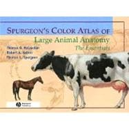
Spurgeon's Color Atlas of Large Animal Anatomy: The Essentials

by ; ;
  • ISBN13:

    9780683306736

  • ISBN10:

    0683306731

  • Edition: 1st
  • Format: Paperback
  • Copyright: 1991-01-16
  • Publisher: Wiley-Blackwell

Note: Supplemental materials are not guaranteed with Rental or Used book purchases.

Purchase Benefits

  • Free Shipping Icon Free Shipping On Orders Over $35!
    Your order must be $35 or more to qualify for free economy shipping. Bulk sales, PO's, Marketplace items, eBooks and apparel do not qualify for this offer.
  • eCampus.com Logo Get Rewarded for Ordering Your Textbooks! Enroll Now
List Price: $93.81 Save up to $28.14
  • Rent Book $65.67
    Add to Cart Free Shipping Icon Free Shipping

    TERM
    PRICE
    DUE
    USUALLY SHIPS IN 3-4 BUSINESS DAYS
    *This item is part of an exclusive publisher rental program and requires an additional convenience fee. This fee will be reflected in the shopping cart.

Supplemental Materials

What is included with this book?

Summary

Extraordinary accuracy and beautiful original artwork are just two features readers will find in this new resource, providing a basic foundation in domestic large animal anatomy. Its unique organization includes the anatomy of all organ systems in the various species, described in a consistent manner. The book presents relevant anatomy of the following species: * Horse (with contributors by Dr. Gayle Trotter) * Ox (with contributions by Dr. Fran Garry) * Sheep and goat (with contributions by Dr. Joan Bowen) * Llama and alpaca (with contributions by Dr. LaRue Johnson) * Swine (with contributions by Dr. LaRue Johnson) * and chicken (with contributions by Dr. John Avens). Features that enhance understanding: * Carefully selected labeling helps students learn and remember structures and relationships. * Male and female of a given species are depicted on facing pages so that topographic anatomy is easy to compare. * Structures common to various animals are labeled several times, whereas unique structrur5es are labeled only on one or two species so students can make rapid distinctions of the structures peculiar to certain animals. * An introduction provides readers with a background in nomenclature and anatomic orientation so they can benefit from the atlas even if they lack training in anatomy. The Atlas depicts topographic relationships of major organs in a simple, yet technically accurate presentation that's free of extraneous detail so that those using the atlas can concentrate on the essential aspects of anatomy.

Author Biography

Thomas O. McCracken and Robert A. Kainer, DVM.

Table of Contents

Introduction xii
Nomenclature and Anatomic Orientation xiii
Animal Classification xiii
General Terminology. Positional and Directional Terms xiv
Body Planes xvi
Body Cavities and Membranes xviii
SECTION 1 THE HORSE (Equus caballus)
Right lateral view of a stallion
2(1)
Left lateral view of a mare
3(1)
Body regions of the horse
4(1)
Skeleton of the horse
5(1)
Cutaneous muscles and major fasciae of the stallion
6(1)
Superficial muscles and veins of the mare
7(1)
A. Parasagittal section of the equine digit
8(1)
Palmar (plantar) view of major structures of the equine digit
8(1)
Relations of the hoof
9(1)
Stay apparatus of the equine forelimb
10(1)
Stay apparatus and reciprocal apparatus of the hindlimb
11(1)
Deep muscles and in situ viscera of the stallion
12(1)
Deep cervical muscles, major joints, and in situ viscera of the mare
13(1)
Median section of the horse's head
14(1)
A. Occlusal (grinding) surfaces of an equine lower first incisor tooth related to continuous eruption and wear
15(1)
B. Complete dentition of the male horse circa 5 years of age
15(1)
Isolated stomach and intestines of the horse
16(1)
Equine cecum, large (ascending) colon, and transverse colon in situ
17(1)
Clinical condition: Right dorsal displacement of the large colon
18(1)
Clinical condition: Left dorsal displacement of the large colon
19(1)
Reproductive organs, urinary organs, liver, heart, and adjacent major vessels related to the skeleton of the stallion
20(1)
Heart and some adjacent major vessels, abdominal and pelvic viscera, and udder (mammary glands) of the mare
21(1)
Relations of the reproductive organs of the stallion
22(1)
Relations of the reproductive organs of the mare
23(1)
Neonatal organs of the foal
24(1)
Major arteries of the mare
25(1)
Major veins of the stallion. Portal system excluded
26(1)
Lymph nodes and vessels of the horse
27(1)
Central and somatic nervous system of the stallion
28(1)
Autonomic nervous system of the mare
29(3)
SECTION 2 THE OX (Bos taurus, also Bos indicus)
Right lateral view of a beef bull
32(1)
Left lateral view of a dairy cow
33(1)
Body regions of the ox
34(1)
Skeleton of the ox
35(1)
Cutaneous muscles and major fasciae of the bull
36(1)
Superficial muscles and veins of the cow
37(1)
Deep cervical muscles and in situ viscera of the bull
38(1)
Deep cervical muscles, major joints, in situ viscera, and udder of the cow
39(1)
Median section of the head and left lateral view of the respiratory system of the ox
40(1)
Interior of the rumen and reticulum of the cow
41(1)
clinical condition: Right volvulus of the abomasum in a bull
42(1)
Clinical condition: Left displacement of the abomasum in a cow
43(1)
Reproductive organs, urinary organs, liver, heart, and adjacent major vessels related to the skeleton of the bull
44(1)
Heart and adjacent major vessels, abdominal and pelvic viscera, and udder (mammary glands) of the cow
45(1)
Relations of the reproductive organs of the bull
46(1)
Relations of the reproductive organs of the cow
47(1)
Major veins of the bull
48(1)
Major arteries of the cow
49(1)
Central nervous system and principal nerves of the peripheral nervous system of the bull
50(1)
Significant lymphatic organs of the cow
51(3)
SECTION 3 THE SHEEP (Ovis aries)
Right lateral view of a ram
54(1)
Left lateral view of an ewe
55(1)
Carcass cuts of the lamb
56(1)
Skeleton of the sheep
57(1)
Cutaneous muscles and major fasciae of the ram
58(1)
Superficial muscles and veins of the ewe
59(1)
Deep cervical muscles and in situ viscera of the ram
60(1)
Deep cervical muscles, in situ viscera, skeleton, and major joints of the ewe
61(1)
Dissection of the parotid region and cross-section of the neck of the sheep
62(1)
A. Location of the left flank incision
63(1)
Cross-section through the left abdominal wall and subjacent ruminal wall
63(1)
Reproductive organs, urinary organs, esophagus and stomach, heart, and adjacent major vessels related to the skeleton of the ram
64(1)
Reproductive organs, urinary organs, heart, and adjacent major vessels, esophagus and stomach of the ewe
65(1)
Relations of the reproductive organs of the ram
66(1)
Relations of the reproductive organs of the ewe
67(1)
Penis of the ram
68(1)
Isolated reproductive organs of the ewe
69(3)
SECTION 4 THE GOAT (Capra hircus)
Right lateral view of an Angora buck (billy)
72(1)
Left lateral view of a Toggenberg doe (nanny)
73(1)
Body regions of the goat
74(1)
Skeleton of the goat
75(1)
Cutaneous muscles and major fasciae of the buck
76(1)
Superficial muscles and veins of the doe
77(1)
Major structures of the caprine left distal metacarpus and digits
78(1)
A. Untrimmed hoofs of the goat
79(1)
Trimmed hoofs of the goat
79(1)
Parasagittal section through the fetlock and digit
79(1)
Deep muscles and in situ viscera of the buck
80(1)
Deep cervical muscles, in situ viscera, skeleton, and major joints of the doe
81(1)
Superficial structures of the goat's head
82(1)
Median section of the caprine head
83(1)
Reproductive organs, abdominal viscera, heart, and adjacent major vessels related to the skeleton of the buck
84(1)
Reproductive organs, abdominal viscera, heart, and adjacent major vessels of the doe
85(1)
Relations of the reproductive organs of the buck
86(1)
Relations of the reproductive organs of the doe
87(3)
SECTION 5 THE LLAMA AND ALPACA (Lama glama and Lama pacos)
Right lateral view of a male llama
90(1)
Left lateral view of a female huacaya alpaca
91(1)
Body regions of the llama
92(1)
Skeleton of the llama
93(1)
Cutaneous muscles and major fasciae of the male llama
94(1)
Superficial muscles of the female alpaca
95(1)
Deep muscles and in situ viscera of the male llama
96(1)
Deep cervical muscles, in situ viscera, and major joints of the female alpaca
97(1)
Major structures of the lamoid left distal metacarpus and digits
98(1)
Median section of the llama's head
99(1)
Proper and improper placement of a halter on a llama's head
100(1)
Relations of the llama's common carotid artery and jugular vein
101(1)
Dentition of the male llama
102(1)
Isolated stomach and intestines of the male llama
103(1)
Reproductive and urinary organs, stomach, liver, heart, and adjacent major vessels related to the skeleton of the male llama
104(1)
Reproductive and urinary organs, stomach, heart, and adjacent major vessels of the female alpaca
105(1)
Relations of the reproductive organs of the male llama
106(1)
Relations of the reproductive organs of the female alpaca
107(3)
SECTION 6 THE SWINE (Sus scrofa domesticus)
Right lateral view of a boar
110(1)
Left lateral view of a sow
111(1)
Carcass cuts of the hog
112(1)
Skeleton of the swine
113(1)
Cutaneous and superficial muscles of the boar
114(1)
Superficial muscles of the sow
115(1)
Deep muscles and in situ viscera of the boar
116(1)
Deep cervical muscles, major joints, and in situ viscera of the sow
117(1)
Median section of the porcine head
118(1)
A. Permanent dentition of the boar
119(1)
Cutting the deciduous incisor and canine teeth of a piglet
119(1)
Isolated stomach and intestines of the swine
120(1)
Lymph nodes and vessels of the sow
121(1)
Reproductive and urinary organs, stomach, liver, heart, and adjacent major vessels related to the skeleton of the boar
122(1)
Reproductive and urinary organs, abdominal viscera, spleen, heart, and adjacent major vessels of the sow
123(1)
Relations of the reproductive organs of the boar
124(1)
Relations of the reproductive organs of the sow
125(3)
SECTION 7 THE CHICKEN (Gallus gallus domesticus)
Right lateral view of a rooster (cock)
128(1)
Left lateral view of a hen
129(1)
Feather coat of the rooster
130(1)
Skeleton of the chicken
131(1)
Superficial muscles of the rooster
132(1)
Superficial muscles of the hen
133(1)
Relations of in situ viscera to the skeleton and cervical muscles of the rooster
134(1)
Relations of in situ viscera and blood vessels to the skeleton and cervical muscles of the hen
135(1)
Isolated gastrointestinal tract of the chicken
136(1)
Air sacs and lungs of the chicken
137(1)
In situ viscera, major blood vessels, and axial skeleton of the rooster
138(1)
In situ viscera, major blood vessels, and axial skeleton of the hen
139(1)
Reproductive organs of the rooster
140(1)
Reproductive organs of the hen
141

Supplemental Materials

What is included with this book?

The New copy of this book will include any supplemental materials advertised. Please check the title of the book to determine if it should include any access cards, study guides, lab manuals, CDs, etc.

The Used, Rental and eBook copies of this book are not guaranteed to include any supplemental materials. Typically, only the book itself is included. This is true even if the title states it includes any access cards, study guides, lab manuals, CDs, etc.

Rewards Program