did-you-know? rent-now

Amazon no longer offers textbook rentals. We do!

did-you-know? rent-now

Amazon no longer offers textbook rentals. We do!

We're the #1 textbook rental company. Let us show you why.

9781608311439

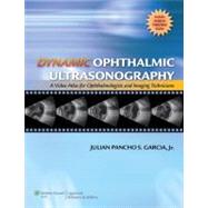
Dynamic Ophthalmic Ultrasonography A Video Atlas for Ophthalmologists and Imaging Technicians (The Advanced Retinal Imaging Center Collection of The New York Eye and Ear Infirmary)

by
  • ISBN13:

    9781608311439

  • ISBN10:

    1608311430

  • Edition: 1st
  • Format: Hardcover
  • Copyright: 2009-09-23
  • Publisher: LWW

Note: Supplemental materials are not guaranteed with Rental or Used book purchases.

Purchase Benefits

List Price: $198.99 Save up to $49.75
  • Buy Used
    $149.24
    Add to Cart Free Shipping Icon Free Shipping

    USUALLY SHIPS IN 2-4 BUSINESS DAYS

Supplemental Materials

What is included with this book?

Summary

The video atlas was written to complement the existing literature on ultrasonography of the eye and orbit. It is a dynamic presentation of various ophthalmic and orbital entities that encountered in a clinical setting. The video atlas is designed to be a quick reference tool in the clinics. The book presents the basic ultrasound movements observed in the eye and orbit. Each ophthalmic tissue demonstrates a specific type of motion unique and inherent to its anatomic structure. This basic movement may change, however, because of inflammatory or degenerative factors, or the presence of other associated findings. Consequently, the motion displayed may no longer be typical for that tissue and may resemble the basic movement of a different anatomic structure, leading to errors in diagnosis. It presents the typical movement of a particular tissue, and then shows the possible types of motion it could manifest in various pathologic situations. By repeatedly watching the movies, one becomes familiar with how ophthalmic tissues behave, and begins to recognize which specific anatomic structures are involved the next time he or she has a similar case. With exposure to the intricacies of ophthalmic tissue dynamics, the reader then learns to interpret ultrasound findings in a more logical manner. Finally, the reader is tested with a number of cases to gauge what has been learned from the previous sections.

Table of Contents

Section I: OVERVIEW Chapter 1: Introduction Introduction How to Obtain Dynamic Ophthalmic Ultrasound Movies Positioning of the Probe Anterior Ocular Segment Imaging Posterior Ocular Segment Imaging Format of the Video Atlas Section II. FORMS OF OPHTHALMIC ULTRASOUND MOVEMENT Chapter 2: Convection Chapter 3: Gravity-Dependent Movement Chapter 4: Reflex Motion Chapter 5: Vascularity External Internal Chapter 6: Aftermovement of Vitreous Chapter 7: Aftermovement of Hyaloid Partial Posterior Vitreous Detachments V-Shaped Posterior Vitreous Detachments Complete Posterior Vitreous Detachments Vitreoschisis Chapter 8: Aftermovement of Retina Focal Retinal Detachments Low-Lying Retinal Detachments Open-Funnel Retinal Detachments Closed-Funnel Retinal Detachments Traction Retinal Detachments Retinoschisis Chapter 9: Aftermovement of Choroid Low-Lying Choroidal Detachments Dome-Shaped Choroidal Detachments Section III: CASE PRESENTATIONS Chapter 10: Complex Dynamic Ultrasound Case Presentations Chapter 11: Limitations of Dynamic Ophthalmic Ultrasonography Bibliography Suggested Readings Appendix: List of Videos Index

Supplemental Materials

What is included with this book?

The New copy of this book will include any supplemental materials advertised. Please check the title of the book to determine if it should include any access cards, study guides, lab manuals, CDs, etc.

The Used, Rental and eBook copies of this book are not guaranteed to include any supplemental materials. Typically, only the book itself is included. This is true even if the title states it includes any access cards, study guides, lab manuals, CDs, etc.

Rewards Program