did-you-know? rent-now

Amazon no longer offers textbook rentals. We do!

did-you-know? rent-now

Amazon no longer offers textbook rentals. We do!

We're the #1 textbook rental company. Let us show you why.

9780849340758

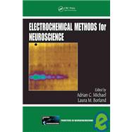
Electrochemical Methods for Neuroscience

by ;
  • ISBN13:

    9780849340758

  • ISBN10:

    0849340756

  • Edition: 1st
  • Format: Hardcover
  • Copyright: 2006-12-13
  • Publisher: CRC Press

Note: Supplemental materials are not guaranteed with Rental or Used book purchases.

Purchase Benefits

  • Free Shipping Icon Free Shipping On Orders Over $35!
    Your order must be $35 or more to qualify for free economy shipping. Bulk sales, PO's, Marketplace items, eBooks and apparel do not qualify for this offer.
  • eCampus.com Logo Get Rewarded for Ordering Your Textbooks! Enroll Now
List Price: $220.00 Save up to $81.40
  • Rent Book $138.60
    Add to Cart Free Shipping Icon Free Shipping

    TERM
    PRICE
    DUE
    USUALLY SHIPS IN 3-5 BUSINESS DAYS
    *This item is part of an exclusive publisher rental program and requires an additional convenience fee. This fee will be reflected in the shopping cart.

Supplemental Materials

What is included with this book?

Summary

Since the first implant of a carbon microelectrode in a rat 35 years ago, there have been substantial advances in the sensitivity, selectivity and temporal resolution of electrochemical techniques. Today, these methods provide neurochemical information that is not accessible by other means. The growing recognition of the versatility of electrochemical techniques indicates a need for a greater understanding of the scientific foundation and use of these powerful tools.Electrochemical Methods for Neuroscience provides an updated summary of the current, albeit evolving, state of the art and lays the scientific foundation for incorporating electrochemical techniques into on-going or newly emerging research programs in the neuroscience disciplines. With contributions from pioneers in the field, the text outlines the applications and benefits of a wide range of electrochemical techniques. It explores the methodology behind the acquisition of neurochemical and neurobiological data through continuous amperometry, fast scan cyclic voltammetry, high-speed chronoamperometry, ion-selective microelectrodes, enzyme based microelectrodes, and in vivo voltammetry with telemetry. The text also introduces emerging concepts in the field such as the correlation of electrochemical recordings with information obtained from patch clamp, electrophysiological, and behavioral techniques.By presenting up-to-date information on the growing collection of electrochemical methods, microsensors, and research techniques, Electrochemical Methods for Neuroscience assists seasoned researchers and newcomers to the field in making sound decisions about adopting the most appropriate of these tools for their future research objectives.

Table of Contents

Chapter 1 An Introduction to Electrochemical Methods in Neuroscience 1
Laura M. Borland and Adrian C. Michael
Introduction
1
A Bit of Electrochemical History: Early Lessons in Selectivity
2
In Vivo Electrochemistry: The Benefits of Size and Speed
3
The Scope of Electrochemistry in the Neurosciences
4
Electrochemistry Fundamentals
7
Chronoamperometry
8
A Comment about Charging Currents
10
Other Potential Step Methods
10
Cyclic Voltammetry
11
Amperometry
12
Conclusion
12
References
13
Chapter 2 Rapid Dopamine Release in Freely Moving Rats 17
Donita L. Robinson and R. Mark Wightman
The Phenomenon of Dopamine Transients
17
Physiology of Dopamine Transients
18
Advances in Recording Technique
19
Measurement of Dopamine Transients: Fast-Scan Cyclic Voltammetry
20
Electrode Preparation
20
Electrode Calibration
21
Surgical Preparation
21
Electrochemical Measurements
22
Instrumentation and Software
22
Experimental Protocol
23
Data Analysis
25
Verification of Recording Site
28
Naturally Occurring Dopamine Transients
28
Environmental Stimuli
28
Basal Conditions
29
Pharmacology
29
Self-Administration of Reinforcers
30
Function of Dopamine Transients
31
References
32
Chapter 3 Presynaptic Regulation of Extracellular Dopamine as Studied by Continuous Amperometry in Anesthetized Animals 35
Marianne Benoit-Marand, Marie-Francoise Suaud-Chagny, and François Gonon
Electrically Evoked Dopamine Release Monitored by Continuous Amperometry
36
Introduction
36
The Quest for Speed
36
Principle of Continuous Amperometry
36
Methods: Carbon Fiber Electrodes and Potentiostats
36
Validity and Field of Application
37
Monitoring Dopamine in the Presence of Other Oxidizable Compounds
37
Amperometric Monitoring of Dopamine Release Evoked by Brief Electrical Stimulation
38
Field of Application
38
Comparison with Other Approaches
39
Comparison with In Vivo Microdialysis
39
Comparison with Fast Scan Cyclic Voltammetry
39
Comparison with Other Electrochemical Techniques
39
Characteristics of Dopaminergic Transmission
40
Dopaminergic Transmission Is Extrasynaptic but Local
40
Release and Elimination of Dopamine from the Extracellular Space
41
Release and Diffusion of Dopamine in the Extracellular Fluid to the Electrode
41
Elimination of Released Dopamine by Reuptake
41
Monitoring the Evoked Dopamine Overflow with Carbon Fiber Electrodes
41
Technical Recommendations
42
Experimental Protocols
42
Carbon Fiber Electrodes
42
Implantation of Carbon Fiber Electrodes In Vivo
43
Reference Electrode
43
In Vitro Calibration of Carbon Fiber Electrodes
43
Analysis of the Data
44
Direct Estimate of Dopamine Release and of Dopamine Uptake
44
Mathematical Models
44
Conclusions
44
References
45
Chapter 4 Fast Scan Cyclic Voltammetry of Dopamine and Serotonin in Mouse Brain Slices 49
Carrie E. John and Sara R. Jones
Introduction
49
Advantages of the Use of Mouse Brain Slices
50
Methodology for Fast Scan Cyclic Voltammetry in Mouse Brain Slices
51
Electrode Fabrication and Calibration
51
FSCV Data Acquisition
52
Brain Slice Preparation
53
Electrically Stimulated Monoamine Release
54
Electrical Stimulation
54
Advantages of the Use of Electrical Stimulation
55
Experimental Design
55
Data Analysis
55
Uptake Rate Determination
55
Regional Variation in Uptake Rates
57
Monoamine Uptake Inhibitors and Releasers
58
Mechanisms of Action
58
Using FSCV to Evaluate Changes in Monoamine Dynamics in Response to Uptake Inhibitors and Releasers
58
Concluding remarks
60
Acknowledgments
60
References
60
Chapter 5 High-Speed Chronoamperometry to Study Kinetics and Mechanisms for Serotonin Clearance In Vivo 63
Lynette C. Daws and Glenn M. Toney
Introduction: Serotonin and the Serotonin Transporter
63
Voltammetric Techniques used to Study Kinetics of Serotonin Clearance in Brain
64
High-Speed Chronoamperometry
64
CFE Calibration
65
High-Speed Chronoamperometry Coupled to Microejection of Serotonin
66
Surgical Procedures
68
Kinetics of Serotonin Clearance
68
Serotonin Signal Parameters
68
Brain Region Dependency
69
Transporter Promiscuity
69
Physiological Relevance of Applying Micromolar Concentrations of 5-HT
73
Biological Significance of Transporter Promiscuity
73
Receptor Regulation of the Serotonin Transporter
74
5-HT 1B Receptor
74
Alpha2-Adrenoceptor
75
Adenosine Receptor
75
Significance of Receptor-Mediated Regulation of 5-HTT
77
Concluding Remarks
77
Acknowledgments
77
References
77
Chapter 6 Using High-Speed Chronoamperometry with Local Dopamine Application to Assess Dopamine Transporter Function 83
Joshua M. Gulley, Gaynor A. Larson, and Nancy R. Zahniser
Introduction
84
Importance of Dopamine and the Dopamine Transporter (DAT)
84
Non-Electrochemical Methods Used to Measure DAT Number and Function
86
Radioligand Binding and Antibodies
86
DA Uptake Assays
86
DAT-Associated Currents
87
In Vivo Microdialysis
87
Electrochemical Methods Used to Measure In Vitro and In Vivo DAT Function
88
Use of High-Speed Chronoamperometry (HSC) with Local DA Application to Measure In Vivo DAT Function in Discrete Brain Regions of Anesthetized Rats and Mice
89
Methods
89
HSC
89
Electrode Construction, Calibration, and Assembly with Micropipettes
89
Surgical Procedures
91
Experimental Details
92
Data Analysis
92
Results
94
Summary of Evidence That This Approach Measures DAT Function
95
Rapid Regulation of DAT Can Influence Measures of Exogenous DA Clearance
95
Additional Applications of HSC with Local DA Application to Measure DAT Function
96
In Vitro Recording in Acutely Prepared Brain Slices
96
In Vivo Chronic Recording in Freely Behaving Rats
97
Future Directions in Using HSC to Measure DAT Function
98
Acknowledgments
98
References
98
Chapter 7 Determining Serotonin and Dopamine Uptake Rates in Synaptosomes Using High-Speed Chronoamperometry 103
Xiomara A. Perez, Amanda J. Bressler, and Anne Milasincic Andrews
Introduction: History of Investigating Monoamine Neurotransmitter Uptake
104
Chronoamperometry Methods for Determining Uptake in Synaptosomes
106
Mouse Models of Altered Uptake
106
Carbon Fiber Microelectrode Construction
107
Preparation of Synaptosomes
107
Chronoamperometry and Electrode Calibration
107
HPLC Analysis of Synaptosomal Filtrates
108
Data Analysis and Statistics
108
Characterization of Microelectrode Responses
109
Effects of Oxygen on Synaptosomal Uptake of Serotonin
109
Effects of Stirring on Synaptosomal Uptake of Serotonin
109
Effects of Uptake Inhibitors on Synaptosomal Uptake of Serotonin
110
Inactivation of Serotonin Transporter Expression Results in a Gene Dose-Dependent Reduction in Serotonin Uptake
111
Uptake Kinetics of Serotonin by Chronoamperometry
111
Chronoamperometry versus Radiochemical Methods: Why Such Discrepancies in Uptake Rates?
113
Determination of Dopamine Uptake in a-Synuclein Transgenic Mice by Chronoamperometry
115
Conclusions
117
Acknowledgments
119
References
119
Chapter 8 Using Fast-Scan Cyclic Voltammetry to Investigate Somatodendritic Dopamine Release 125
Sarah Threlfell and Stephanie J. Cragg
What Is Somatodendritic Dopamine Release?
126
Midbrain Dopamine Neurons Release Dopamine from Their Somatodendrites
126
The Functions and Sites of Action of Somatodendritic Dopamine
126
Methodological Approaches to the Study of Somatodendritic Dopamine Release
126
Electrochemical and Other Approaches to Dopamine Detection
126
Co-Detection of DA and 5-HT in Midbrain
127
FCV Waveform Choice
127
Species Variation in 5-HT Interference
129
Characteristic Voltammetric and Pharmacological Differences
130
In Vitro Methods for FCV in Midbrain
130
Slice Preparations and Species
130
Solutions
131
Scan Waveforms
131
Carbon-Fiber Microelectrodes: Preparation, Calibration
133
Choice of Stimulation Parameters
133
Concerns in Signal Identification
135
What Is the Mechanism of Release?
136
Anatomical Evidence
136
Mechanistic Evidence
136
Regulation of Somatodendritic Dopamine Signals by Extracellular Geometry and Uptake
137
Volume Transmission and Uptake
137
Regional Kinetic Distinctions
138
Neuromodulation of Somatodendritic Release
139
Autoreceptor Regulation
139
Heteroreceptor and Other Neuromodulatory Regulation
140
Summary and Conclusions
141
Acknowledgments
142
References
142
Chapter 9 From Interferant Anion to Neuromodulator: Ascorbate Oxidizes Its Way to Respectability 149
George V. Rebec
Introduction
149
Basic Chemistry
150
Isolation and Tissue Availability
150
Oxidation
151
Ascorbate Electrochemistry
151
Methodology
152
Extracellular Changes
155
Glutamate-Related Mechanisms of Striatal Ascorbate Release
156
Ascorbate Modulation of Glutamate-Evoked Neuronal Signaling
157
Ascorbate and Huntington's Disease
158
HD and Transgenic Mice
158
Ascorbate Dysregulation in HD Striatum
158
Summary and Conclusion
161
Acknowledgments
161
References
161
Chapter 10 Biophysical Properties of Brain Extracellular Space Explored with Ion-Selective Microelectrodes, Integrative Optical Imaging and Related Techniques. 167
Sabina Hrabetová and Charles Nicholson
Introduction: Extracellular Space
168
Brief History
169
Volume Fraction and Width
169
Hindrance and Tortuosity
170
Bulk Flow
170
Volume Transmission and Drug Delivery
170
Tools and Methods to Study Extracellular Space in Real Time
171
Diffusion Measurements Reveal Extracellular Space Properties
171
The Diffusion Equation and Its Meaning
171
Choice of Molecular Probes for Extracellular Space
173
Delivery of Molecular Probes: Iontophoresis and Pressure Ejection
175
Iontophoresis
176
Pressure Ejection
178
Detection of Molecular Probes: Ion-Selective Microelectrodes, Integrative Optical Imaging and Carbon-Based Microelectrodes
179
Ion-Selective Microelectrodes
179
Integrative Optical Imaging
181
Carbon-Based Microelectrodes
182
Diffusion Measurements Using Dual Microdialysis Probes
184
Choice of Brain Tissue Preparation
185
Tissue Slices and Slabs
185
Effects of Boundary Conditions on Diffusion Measurements in Brain Tissue
185
In Vivo Preparations
190
Specialized Software for Point-Source Diffusion Analysis
191
Software for Intophoresis and Pressure Ejection-VOLTORO, Walter and Wanda
191
Software for Integrative Optical Imaging Vida and Ida
191
Diffusion Properties of Brain Extracellular Space
192
Brain under Physiological Conditions
192
Brain during Reversible Osmotic Challenge
193
Brain in Pathological States
194
Monte Carlo Simulation of Diffusion in 3D Media
194
MCell and DReAMM
195
Three-Dimensional Media Composed of Convex Cells
195
Dwell-Time Diffusion Theory
196
Three-Dimensional Media Containing Concave Cells and Lakes
197
Future Directions
198
References
199
Chapter 11 Hydrogen Peroxide as a Diffusible Messenger: Evidence from Voltammetric Studies of Dopamine Release in Brain Slices 205
Margaret E. Rice, Marat V. Avshalumov, and Jyoti C. Patel
Introduction
206
Overview
206
Physiology of DA Release: Why It Can Be Detected in the Extracellular Space
206
A Brief History of In Vivo and In Vitro Voltammetry
208
Advantages of Brain Slices to Study DA Release
209
Voltammetric Methods
210
Comparison of Voltammetric Techniques
210
Constant Potential Amperometry
211
Chronoamperometry
211
Fast-Scan Cyclic Voltammetry
212
Electrodes
214
Brain Slice Methodology
214
aCSF Composition for Healthy Slices
216
Species Selection
217
Plane of Slicing
217
Stimulating and Recording DA Release in Brain Slices
219
Signal versus Interference
220
Regulation of Striatal DA Release by H2O2
221
Endogenous H2O2 Inhibits Axonal DA Release
222
Potential Sources of H2O2 Generation
222
Regulation of Axonal DA Release by Glutamate Acting at AMPA Receptors Requires H2O2
222
Activity-Dependent H2O2 Generation in Striatal Medium Spiny Neurons
224
H2O2 Acts via KATp Channels to Inhibit DA Release
225
Summary and Future Directions for Studies of H2O2 as a Neuromodulator
225
Acknowledgments
226
References
226
Chapter 12 In Vivo Voltammetry with Telemetry 233
Paul A. Garris, Phillip G. Greco, Stefan G. Sandberg, Greg Howes, Sirinun Pongmaytegul, Byron A. Heidenreich, Joseph M. Casto, Robert Ensman, John Poehlman, Andy Alexander, and George V. Rebec
Introduction
234
Overview
234
Monitoring Neural Activity
234
Voltammetry
236
Principles of FSCV at a CFM
236
Applications of FSCV at a CFM
238
Principles of Telemetry
240
Resources
240
Theory and Basic Instrumentation
240
Digital Telemetry at 2.4 GHz
242
Wireless Neural Monitoring and Control
244
Scope
244
Electrophysiology
244
Voltammetry
245
Electrical Stimulation
245
Real-Time Animal Telemetry
245
Instrument Overview
245
Establishment of Proof of Principle
247
Preliminary Findings in Freely Moving Animals
248
Future Directions
250
The Smaller the Better
250
Multifunctional Wireless Instrument
250
Portable Microsensor Measurement System
251
Conclusions
253
Acknowledgments
253
References
253
Chapter 13 Oxidative Stress at the Single Cell Level 261
Christian Amatore and Stéphane Arbault
Introduction
261
Oxidative Stress Processes
261
Electroactive Species Implicated in Oxidative Stress
265
Direct Electrochemical Detection of Oxidative Release on Single Cells
266
Microelectrode Detection of Superoxide and Hydrogen Peroxide
Released by Living Cells
268
Detection of Superoxide by Carbon and Gold Microelectrodes on Phagocytes and Vascular Cells
268
Detection of Superoxide by Cytochrome c Modified Gold Electrodes on Neuronal Cells
270
Detection of Hydrogen Peroxide by Platinized Carbon Fiber Microelectrodes on Skin Fibroblasts
272
Microelectrode Detection of Nitric Oxide and Peroxynitrite Releases by Living Cells
273
Detection of Nitric Oxide by Porphyrin Modified Microelectrodes at Single Cells from Cardiac Endothelium
273
Detection of Peroxynitrite by Modified Microelectrodes during Ischemia of Endothelial Cells from Heart
275
Detection of Peroxynitrite by Platinized Microelectrodes on Single Human Fibroblasts
276
Conclusions and Perspectives
278
References
279
Chapter 14 Electrochemistry at the Cell Membrane/Solution Interface 285
Nathan Wittenberg, Marc Maxson, Daniel Eves, Ann-Sofie Cans, and Andrew G. Ewing
Electrochemistry at the Cell Membrane/Solution Interface
286
Basics of Electrodes for Single Cell Measurements
286
Electrode Fabrication and Testing
286
Basic Amperometry
287
Exocytosis at Adrenal Cells
289
Amperometric Detection of Release via the Fusion Pore
289
Insulin Release from Pancreatic Beta Cells
290
Exocytosis at Mast Cells
291
Pheochromocytoma (PC12) Cells, Undifferentiated and Differentiated
291
Release from the Cell Body of a Processed Neuron in an Animal System
291
Release of Small Amounts of Catecholamine from the Leech Neuron
292
Release from Mammalian Cells
292
Kiss-and-Run Measurements
292
Measurements of Cholesterol in the Cell Membrane
292
Other Techniques
292
Patch Amperometry
292
Quartz Crystal Microbalance Measurements
294
Electrochemistry of Material Released from Cells in Microvials
295
Electrochemistry at Bilayer Membranes
297
Artificial Cells (Liposomes) as Models of Exocytosis
297
Kinetics of Membrane Distention during Artificial Cell Exocytosis
297
Modeling Release via the Fusion Pore (Lipid Nanotube)
299
Artificial Synapses with Liposome Models
301
Model of the Synapse
301
Quantitative Issues and Flow Effects in the Synapse
303
Electrochemical Methods Applied to Neuroscience
305
Stages of Release during Exocytosis Elucidated with Amperometry
305
Theories on the Forces Involved in Opening the Fusion Pore
306
Effects of Snare Protein Manipulation
306
Blocking Ryanodine Channels Affects Exocytosis
307
Regulation of Insulin Exocytosis from Pancreatic Beta Cells
307
Vesicle Size Changes with Messenger Amount
308
Summary and Future Perspectives
308
Acknowledgments
308
References
309
Chapter 15 The Patch Amperometry Technique: Design of a Method to Study Exocytosis of Single Vesicles 315
Gregor Dernick, Guillermo Alvarez de Toledo, and Manfred Lindau
Introduction: Capacitance Measurements of Membrane Fusion with Amperometric Detection of Released Molecules
316
Patch Amperometry
317
Patch Amperometry Requires Reversed Patch Clamp Electrode Configuration
317
Equipment and Setup
317
Data Acquisition
317
Capacitance Measurements
318
Amperomety
322
Microscopy
322
Design of the Electrode Holders
322
Manually Adjustable Electrode Holder
323
Motorized Electrode Holder
324
Patch Pipettes
326
Carbon Fiber Electrode Fabrication
326
Carbon Fiber Fabrication Setup
326
Carbon Fiber Pulling
327
Testing the CFE in the Patch Amperometry Configuration
328
Recording Chamber Design
329
Patching of Chromaffin Cells
329
Capacitance Calibration
330
Analysis of Exocytotic Events
331
Vesicle Capactiance and Fusion Pore Conductance
331
Release of Molecules
333
Quantal Size, Vesicle Size, and the Fusion Pore
334
Summary and Discussion
334
Acknowledgments
334
References
335
Chapter 16 Amperometric Detection of Dopamine Exocytosis from Synaptic Terminals 337
Roland G.W. Staal, Stephen Rayport, and David Sulzer
Introduction to Dopamine Neurotransmission
337
Electrochemical Detection of DA
338
Quantal DA Release
338
Source of DA Neurons: Selection of Animals and Brain Regions
339
Methods
340
Preparation of Glass Coverslips
341
Preparation of Pipette "Tech-Tips" for Triturating Cells
342
SYLGARD Circles
342
Glia (Using Rat Pups)
342
DA Neurons
343
Media and Reagents
349
Carbon Fiber Amprometry
349
Electrode Fabrication
349
Testing and Selection of Electrodes
350
Recording
350
Analysis
351
References
351
Chapter 17 Scanning Electrochemical Microscopy as a Tool in Neuroscience 353
Albert Schulte and Wolfgang Schuhmann
Introduction
353
Operational Principles of the Amperometric Feedback and the Generator/Collector Mode of SECM
356
Constant-Height Mode SECM Measurement at Individual Cells
359
Constant-Distance Mode SECM at Individual Cells and Cell Populations
363
Conclusion and Future Aspects
366
References
367
Chapter 18 Principles, Development and Applications of Self-Referencing Electrochemical Microelectrodes to the Determination of Fluxes at Cell Membranes 373
Peter J.S. Smith, Richard H. Sanger, and Mark A. Messerli
Introduction
374
Self-Referencing: Principles
376
Self-Referencing: Potentiometric Ion-Selective Electrodes (ISE)
378
Types of Potentiometric Ion-Selective Microelectrodes
381
Choosing an Electrolyte
383
Self-Referencing: Amperometric Microelectrodes
383
Types of Amperometric Microelectrodes
384
Calculation of Flux
384
Setting the Frequency and Distance of Translation
385
Electrode Construction
385
Electrode Construction: Potentiometric
386
Electrode Construction: Amperometric
388
Making an Oxygen Electrode
388
Making a Hydrogen Peroxide Electrode
388
Making an Ascorbate Electrode
389
Making a Nitric Oxide Electrode
389
Making a Glucose (Enzyme-Assisted) Electrode
390
Making a Glutamate Electrode
390
Response Times
390
Response Times: ISEs
391
Response Times: Amperometric
391
Response Times: Interferents
392
Spatial Resolution
392
Problems and Pitfalls
393
Potentiometric: Selectivity
393
Amperometric: Selectivity
393
Mixing
393
Positional Artifacts
394
Positional Artifacts: Potentiometric
394
Positional Artifacts: Amperometric
394
Voltage Fields
394
Use of Buffers
395
Equipment and Software
396
Preamplifiers
396
Voltage Head Stage Potentiometric
396
Current Head Stage-Amperometric
396
Amplifier
396
Motion Control
397
Software
397
Applications
397
Neural Repair and Disease
397
Microglia
397
Neural Tube Defects
398
Sensory Neurobiology
398
Hair Cells
398
Vision
398
Olfaction
398
Additional Electrically Active Cells
399
Neurons
399
Muscle
399
Pancreatic 13-cells
399
Additional Pumps and Channels
400
Apoptosis and Volume Changes
400
Acidification and Alkalinization
400
Conclusions
401
Acknowledgments
401
References
401
Chapter 19 Second-by-Second Measures of L-Glutamate and Other Neurotransmitters Using Enzyme-Based Microelectrode Arrays 407
Kevin N. Hascup, Erin C. Rutherford, Jorge E. Quintero, B. Keith Day, Justin R. Nickell, Francois Pomerleau, Peter Huettl, Jason J. Burmeister, and Greg A. Gerhardt
Introduction
408
Principles of In Vivo Electrochemistry
408
Enzyme-Based Multisite Microelectrode Arrays
409
Fabrication
409
Multisite Microelectrode Array Designs
412
Headstage/Recording System
413
Microelectrode Preparation
413
Cleaning Procedures
414
Exclusion Layer Coatings
414
Nafion Exclusion Layer
414
1,3-Phenylenediamine
415
Enzyme Layer Coatings
416
Oxidase Enzymes
416
L-Glutamate Oxidase Coating Procedure
416
Calibration
418
Calibration Preparation
418
Calibration Procedures
419
Microelectrode Calibration Criteria
419
Glass Micropipettes for Local Drug Delivery
421
Ag/AgCl Reference Electrode
423
Signal Analysis
423
Amperometric Recordings Utilizing Self-Referencing Microelectrodes
425
Importance of Self-Referencing Microelectrode Arrays
425
Phasic Release of Neurotransmitters
427
Tonic or Basal Release of Neurotransmitters
428
Application to Biological Systems
429
In Vitro Systems
429
Cell Culture Techniques
429
Brain Slice Techniques
430
In Vivo Anesthetized Animal Recordings
433
Anesthetized Rats and Mice
433
Anesthetized Rhesus Monkeys
435
Awake, Freely Moving Rats and Mice
437
Implantation and Recording Procedure
439
Chronic Implantation Histopathology
443
Future Directions
443
Eight Site Microelectrode Arrays
443
Using Microelectrode Microdrives for Chronic Implantations
445
Telemetry
446
Deep Brain Structure Enzyme-Based Microelectrodes
446
Acknowledgments
446
Appendix A Preparing Glutamate Microelectrodes
447
References
448
Chapter 20 Telemetry for Biosensor Systems 451
David A. Johnson and George S. Wilson
Introduction
451
Fast Scan Cyclic Voltammetry
452
FSCV System Requirements
452
In-Vivo Electrochemical Sensors
453
Biosensor System Requirements
454
General Design Considerations
454
Electronics
455
Power Sources
455
RF Telemetry
458
Protocol Independent Transmitters and Receivers
458
ZigBee (IEEE 802.15.4)
458
Bluetooth
459
WiFi (IEEE 802.11)
459
Ultra Wideband
459
Radio Frequency Identification
459
Interference
460
Mesh Networks
460
Antenna Design
461
IR Telemetry
461
Design Examples
461
FSCV
461
Selective Amperometric Biosensors
462
Conclusions
462
References
462
Chapter 21 The Principles, Development and Application of Microelectrodes for the In Vivo Determination of Nitric Oxide 465
Michael J. Serpe and Xueji Zhang
Introduction
465
NO Microelectrodes
466
Clark Type NO Microelectrodes
467
Metalloporphyrin and Metallophthalocyanine Modified NO Microelectrodes
468
Combination NO Microelectrodes
469
Other NO Microelectrodes
470
Calibration of NO Microelectrodes
470
Calibration Based on Chemical Generation of NO
470
Calibration Using an NO Standard Solution
471
Calibration Based on Decomposition of SNAP
471
Characterization of NO Microelectrodes
473
Sensitivity and Detection Limit
473
Selectivity
473
Response Time
474
Selected NO Microelectrode Applications
474
Concluding Remarks
481
Acknowledgments
481
References
482
Chapter 22 In Vivo Fast-Scan Cyclic Voltammetry of Dopamine near Microdialysis Probes 489
Hua Yang and Adrian C. Michael
Introduction
489
Procedures for Voltammetry near Microdialysis Probes
491
Voltammetric Electrodes and Procedures
491
Dialytrode Design
491
Surgical Procedures
491
Dual Electrode Voltammetry
492
Voltammetry near Microdialysis Probes
492
Results
492
Dual Electrode Recording
492
Probe Implantation Affects the Voltammetric Responses
494
Discussion and Implications
496
Acknowledgments
499
References
500
Index 503

Supplemental Materials

What is included with this book?

The New copy of this book will include any supplemental materials advertised. Please check the title of the book to determine if it should include any access cards, study guides, lab manuals, CDs, etc.

The Used, Rental and eBook copies of this book are not guaranteed to include any supplemental materials. Typically, only the book itself is included. This is true even if the title states it includes any access cards, study guides, lab manuals, CDs, etc.

Rewards Program