did-you-know? rent-now

Amazon no longer offers textbook rentals. We do!

did-you-know? rent-now

Amazon no longer offers textbook rentals. We do!

We're the #1 textbook rental company. Let us show you why.

9780345457110

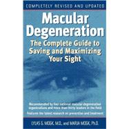
Macular Degeneration The Complete Guide to Saving and Maximizing Your Sight

by ;
  • ISBN13:

    9780345457110

  • ISBN10:

    0345457110

  • Format: Paperback
  • Copyright: 2003-01-01
  • Publisher: Ballantine Books

Note: Supplemental materials are not guaranteed with Rental or Used book purchases.

Purchase Benefits

  • Free Shipping Icon Free Shipping On Orders Over $35!
    Your order must be $35 or more to qualify for free economy shipping. Bulk sales, PO's, Marketplace items, eBooks and apparel do not qualify for this offer.
  • eCampus.com Logo Get Rewarded for Ordering Your Textbooks! Enroll Now
List Price: $18.00 Save up to $4.50
  • Buy Used
    $13.50

    USUALLY SHIPS IN 2-4 BUSINESS DAYS

Supplemental Materials

What is included with this book?

Summary

The acclaimed book on macular degenerationnow completely revised and updated with cutting edge research and the latest developments in the field. More than fifteen million Americans have age-related macular degeneration (AMD), and the disease will strike 200,000 more people this year. It is the most prevalent cause of vision loss in the western world. Dr. Lylas Mogk, the founding director of the Visual Rehabilitation and Research Center of the Henry Ford Health System, has a unique professional and personal understanding of AMD. A doctor and loving daughter of a parent with this frightening though manageable condition, Mogk here explains exactly what it is and how to limit its effect on your life. Reassuring and comprehensivecomplete with illuminating first person stories of people with AMDMacular Degenerationwill help you or someone you love with information on Reducing your risk factors Revolutionary new technology, including laser surgery and alternative treatments New research discoveries in nutritionand eye-healthy recipes The latest low-vision computer software programs Coping with depression and frustration Active online communities of people with macular degeneration Plusa Low Vision Living Rehab program to help you read better, see better, and live independently!

Author Biography

Lylas G. Mogk, M.D., is a practicing ophthalmologist and founding Director of the Visual Rehabilitation and Research Center of Michigan, part of the Henry Ford Health System. Dr. Mogk is a member of the American Academy of Ophthalmology's Low Vision Rehabilitation Committee. She lives in Grosse Pointe with her husband, John, and her father, Charles R. Good, who has advanced macular degeneration. Despite his severe vision loss, Dr. Mogk's father lives a full and active life following the principles presented in this book.<br><br>Marja Mogk is a freelance writer and editor based in Berkeley, California. She worked for several years in social welfare counseling and maintains an active interest in this field. She is Lylas and John Mogk's daughter.

Table of Contents

Acknowledgmentsp. xix
Introduction: My Family's Storyp. 1
Understanding AMD
What Is AMD? A Portraitp. 9
Medical Treatments, Research, and New Discoveriesp. 42
Genes, Greens, and Oils: The Causes, Prevention, and Natural Treatment of AMDp. 88
New Gourmet Greens: Recipes for Your Eyesp. 132
Experiencing AMD
I Am Not Blind: The Shock of AMDp. 155
With the Heart One Sees Rightly: Living Fully with AMDp. 182
Sixteen Tips for Family and Friendsp. 225
I See Purple Flowers Everywhere: The Many Visions of Charles Bonnet Syndromep. 236
Addressing AMD
Why Visual Rehabilitation Is Right for Youp. 255
Making Things Brighter: Lightingp. 264
Your Reading Workshopp. 276
Making Things Bigger: Magnifiers, Telescopes, and Computersp. 298
Saving Sight on the Road: Driving, Alternatives, and Travelingp. 337
Saving Sight at Home and in Your Communityp. 362
Appendixes and Index
Macular Degeneration Organizations, Finding Visual Rehabilitation Programs in Your Area, On-line Communities, and Memoirs by People with Vision Lossp. 403
Reading, Watching, and Listeningp. 410
The Best Low-Vision Product Catalogs, and CCTVs, Computers, and Softwarep. 422
Driving, Civil Rights, Tax Benefits, and Donations and Bequestsp. 429
Research Updates and Nutrition Studiesp. 434
Canada and Canadians with AMDp. 436
Indexp. 441
About the Authorsp. 457
Table of Contents provided by Rittenhouse. All Rights Reserved.

Supplemental Materials

What is included with this book?

The New copy of this book will include any supplemental materials advertised. Please check the title of the book to determine if it should include any access cards, study guides, lab manuals, CDs, etc.

The Used, Rental and eBook copies of this book are not guaranteed to include any supplemental materials. Typically, only the book itself is included. This is true even if the title states it includes any access cards, study guides, lab manuals, CDs, etc.

Excerpts

What Is AMD?
A Portrait

This has become a true epidemic of our time.

–Jerry Chader, Ph.D.
Chief Scientific Officer
The Foundation Fighting Blindness

Ten years ago, when Zelda Grant discovered she had age-related macular degeneration, she was astounded. “Macular what?” she said. She had never heard of it before. Today most of us know something about AMD, but it still comes as a shock. “I couldn’t believe it,” Edina Williams told me, “I kept thinking: I need new glasses. A new pair will solve the problem. I still can’t believe it.” Age-related macular degeneration, or AMD, is the leading cause of adult vision loss in North America, Europe, and Australia. It affects the sight of more adults than all of the better-known eye diseases combined: glaucoma, cataracts, and diabetic retinopathy. According to the Beaver Dam Eye Study, 18 percent of seniors aged sixty-five to seventy-four and nearly 30 percent of seniors over seventy-five show early evidence of the condi- tion. One out of every twenty-five Americans over sixty-five, or nearly 1.5 million people, have already experienced significant vision loss from advanced macular degeneration, and 200,000 more Americans lose vision every year. Hundreds of thousands of people in Canada, England, Germany, and many other countries have lost vision from AMD, too. Clearly, if you have AMD you are not alone.

What Is Age-Related
Macular Degeneration?

Macular degeneration dismantles central vision painlessly and silently, leaving peripheral vision intact. As a result, people with advanced macular degeneration do not feel any discomfort in their eyes and they do not appear any different to
their friends and family, but their experience of the world and of their own capacities changes radically.

Because macular degeneration leaves peripheral vision intact, people with AMD can see whatever rests at the edges of their vision but cannot see clearly whatever they look at directly. They find it difficult to recognize their grandchildren’s photographs, for example, but can describe the check pattern of a black-and-white tile floor. They cannot read a bus sign but can see a green leaf on the sidewalk out of the corners of their eyes. This combination of visual ability and vision loss is enormously frustrating, not the least because it takes away what we most want to see, leaving visible what appears to be less important. As Carolyn See remarked dryly in her candid article in Modern Maturity about living with macular degeneration, “It begins in the center of your vision and after a while you can’t read or drive or recognize your relatives. They say you’ll always be able to pick up a thread on the carpet. But even with full vision, picking up threads on the carpet wasn’t high on my list of activities.”

Why Has Macular Degeneration Suddenly Become So Common?

“Ten years ago, when I met someone on a plane en route to giving a talk and told them what I
do, they would look at me kinda funny,” reflected Nikolai Stevenson, president of the Association for Macular Diseases, an organization run for and by people with AMD. “With a name like ‘macular degeneration,’ for all they knew it was contagious. Today, though, everyone seems to know someone else with it, too: a neighbor, a friend, or a relative.” Macular degeneration is no longer a stealth intruder but a known one. Why is AMD suddenly so common? Is this a new epidemic?

Doctors have actually known about macular degeneration for more than 100 years. AMD was first named by a German scientist in 1885, but the technology used to see it clearly was not developed until the 1960s. Unlike cataracts, which are visible to the naked eye, or glaucoma, which can easily be measured, macular degeneration was historically more difficult to detect, analyze, and treat. As a result, other eye conditions that were easier to understand and responded well to surgery and medication received much more attention from the medical community during the twentieth century. Macular degeneration fell to the bottom of the priority list, until now.

Are more people getting it today than ever before? Yes, but we aren’t exactly sure why. On the one hand, it may be a result of our eating habits, food production methods, and the effects of more than half a century of heavy-duty industrial pollution (see chapter 3 for a discussion of the causes of macular degeneration and what you can do to minimize your risk). On the other hand, there are simply more of us today and we are living longer and healthier lives than ever before. The longer you live, the more likely you are to develop age-related macular degeneration, which doesn’t usually affect people until they pass the age of fifty and increases exponentially every decade thereafter (hence the prefix age-related).

It’s also true that many of our parents or grandparents may have had macular degeneration, but we didn’t recognize their vision loss as a disease. We saw it simply as a sign of growing old. I remember my own grandmother stooped over in the garden wearing her black dress and black shoes, with her gray hair tucked in a bun. It was 1947 and she was only sixty-nine, but we thought she was very, very, very old. We also knew she couldn’t see well, but no one had a special name for that. Today sixty-nine is looking pretty young. The folks in their late sixties I see at our visual rehabilitation program in Michigan come in wearing very spiffy outfits and have a good fifteen to thirty more good years of reading, entertaining, traveling, and sports ahead of them. Some of them aren’t even retired. In fact, sometimes I see ninety-year-olds who fit that description. If I suggested that they wear black, put their hair in a bun, and act old like my grandma was, they’d probably slug me. Seniors today aren’t old the way they used to be, and they aren’t willing
to pass off age-related macular degeneration as something that “just happens” when you’re “old,” nor should they.

Macular Degeneration Gains Attention

In the last ten years, macular degeneration has become a major health issue. The National Eye Institute (NEI), a division of the National Institutes of Health, has made AMD its number-one priority, and Medicare has approved national coverage for visual rehabilitation for people with vision loss. Studies are underway at major research centers across the country to discover the genetic components of AMD and its environmental causes and to find a cure. Many new surg- eries and treatments are on the horizon. Chap- ter 2, “Medical Treatments, Research, and New Discoveries,” outlines all of the exciting new developments in the field. For the latest research updates, see the resources listed in appendix E. We now know for sure that nutrition can help prevent the disease: what you eat does make a difference. See chapter 3, “Genes, Greens, and Oils: The Causes, Prevention, and Natural Treatment of AMD,” for details and diet recommendations. Macular degeneration is complicated, and so far there is no single easy answer–but we do have some answers and we have a very strong hope that many more are soon to come.

Macular Degeneration Isn’t
Just About Your Eyes

Most people say they’d rather lose a limb than
an eye, and national surveys tend to place vision loss among the most feared afflictions, along with Alzheimer’s. Why? Because eyesight affects every aspect of life: mobility, physical activity, communication, appearance, perception, self-esteem, and psychological health. Macular degeneration is not just about how much you can or cannot see. It’s about your whole life: how you cope with change, your view of the future, and your capacity to enjoy the present. And macular degeneration is tailor-made to push every button we have. It can raise feelings of grief, helplessness, depression, fear, anxiety, and anger. Part II of this book addresses these experiences and how you can live fully with AMD, but first we have to really understand the condition itself. As Zelda put it, “Macular what?” What is this macular degeneration really?

AGE-RELATED MACULAR
DEGENERATION EXPLAINED

Our Eyes Are Like Little Cameras

You have probably heard this analogy before: Our eyes are like little cameras. Just as light enters the camera through the shutter, is focused
by the lens, and falls on the film, so light enters
our eye through the pupil, is focused by the lens, and falls on the retina at the back of our eye.
The retina is like camera film. Its thin tissue forms the inner lining of the eye, picking up light and converting it into nerve signals. The retina sends those signals through the optic nerve to
the brain, which “develops” them into the images we actually see, just as film is developed into photographs.

The Macula: Center of the Retina

The retina has two types of photoreceptor cells that convert light into electrical messages for the optic nerve to transmit: rod cells and cone cells, so named for their shapes. There are many more rods than cones throughout the retina, especially at the edges, where rods outnumber cones twenty to one. Rod cells are responsible for light-and-dark contrast perception. They essentially provide us with background information, but they cannot transmit crisp pictures. We use the rods of our peripheral vision to catch a glimpse of something. They tell us that a car is coming from the far left or right, but in order to see the car clearly or describe it, we instinctively turn to look at it directly. As soon as we turn, however, we are
no longer primarily using our rod cells to see but our cone cells. Cones are concentrated in the center of the retina–called the macula–and are responsible for central vision, color perception, and sharp images (acute vision). The capacity of cones to distinguish detail is one hundred times greater than that of rods. We need them to tell the difference between forest green and black and to
see precise detail, such as the features of a face, the lace pattern on a tablecloth, and the letters on this page. The macula is therefore both the geographic center of the retina and the focal center of our vision. The fovea is the very center of the macula. It is also the only area of the retina that has only cone cells. For all its power, though, the macula is very small; it measures about a quarter inch in diameter and is tissue paper thin. But the macula is truly a mouse that roars. This tiny area is responsible for so much of what we see.

Macular Degeneration: The Key Players

In macular degeneration, rod and cone cells of the macula begin to die, reducing the number of cells able to transmit visual signals to the brain. Macular degeneration, however, is not a condition of these cells alone but also of the underlying tissue that supports them and keeps them healthy. In addition to the rods and cones, there are three more key players in macular degeneration: the retinal pigment epithelium (RPE), Bruch’s membrane, and the choroid. In that order, each is a layer of tissue that lies beneath the retina, like layers of a club sandwich or, more accurately, stations on a delivery line. Taken together, they form a kind of conveyor belt for nutrition and waste management, constantly supplying the macula with oxygen-laden meals and whisking away waste. The large blood vessels of the choroid truck materials in and out through the bloodstream. Bruch’s membrane acts as a security gate between these blood vessels and the delicate RPE, and the RPE delivers oxygen and receives waste directly from the rod and cone cells in the macula.

Normally, the system works very efficiently. But if there’s a jam somewhere, the oxygen
meal shipments and the waste removals back
up, clogging the pickup and drop-off stations
until they eventually shut down. The choroid, Bruch’s membrane, and the RPE become disabled and can no longer do their jobs. When they fail, the rod and cone cells lack the massive amounts of oxygen they need to stay alive and cannot clear away the waste products they produce. Dying of oxygen deprivation and clogged with refuse, rods and cones become unable to send signals through the optic nerve to the brain–they are no longer able to see. This is what happens with macular degeneration.

TWO TYPES OF AMD:
DRY AND WET

There are two types of macular degeneration, commonly called dry and wet. All cases are thought to start with the dry form. Between 10 and 15 percent of the people who show signs of dry macular degeneration eventually develop the wet form.

Dry AMD

Although there is only one kind of dry AMD, you may hear it called atrophic, geographic atrophy, or nonexudative macular degeneration. Atrophic or atrophy refers to a declining, weakening, or wasting away. We often use the word to talk about muscles that haven’t been used in a great while and lose their strength as a result. We can exercise our muscles and regain strength, but unlike our muscles, atrophy in our macula isn’t currently reversible. This is because the macula atrophies from a lack of oxygen, not a lack of use. And, as we know, any part of our body that suffers a prolonged lack of oxygen usually sustains permanent damage. Geographic atrophy of the macula means atrophy concentrated in one contiguous area of the macula. Nonexudative means not exuding, or not
discharging: there is no blood leakage in the macula, contributing to the malfunctioning of the conveyor belt system.

HARD AND SOFT DRUSEN

Dry AMD is usually signaled by the presence in the macula of small pale spots called drusen. There are two type of drusen: less harmful hard drusen and more ominous soft drusen. Hard drusen are small, round, sharply defined light yellow deposits of lipid (a fatty compound) and calcium that accumulate on Bruch’s membrane. They are quite common with age, appearing in most older eyes like age spots appear on skin, and are not necessarily thought to indicate macular degenera- tion. Soft drusen can be nearly twice the size of hard drusen, with indistinct margins and varying sizes and shapes. While soft drusen can be seen in older eyes that don’t develop full-blown AMD, they have been considered an early indicator of the condition, perhaps because they are the first feature of AMD that we can detect in an affected eye. Recently, however, researchers have suggested that by the time we can see soft drusen in an eye, macular degeneration may already be advanced.

Soft drusen are thought to plug up the conveyor belt system in dry macular degeneration. Some researchers also believe that soft drusen are responsible for wet macular degeneration because they may weaken Bruch’s membrane or because they may trigger the proliferation of abnormal blood vessels. Other researchers disagree, arguing that soft drusen occur because Bruch’s membrane has already been weakened for some other reason. In any case, soft drusen signal to us that the conveyor belt support system for the macula is malfunctioning and Bruch’s membrane is weak, which may allow abnormal blood vessels from the choroid to creep through.

FOCAL HYPERPIGMENTATION

Like both hard and soft drusen, focal hyperpigmentation is a signal of possible early macular degeneration that your ophthalmologist can see during a standard dilated eye exam with no special testing. Focal hyperpigmentation means the appearance of darkish irregular specks in the macula. These specks are caused by pigment cells that clump up over time, although we aren’t sure exactly why they do. Although the retina is transparent, it appears to us as reddish-orange because the underlying RPE and choroid give it color. The light yellow color of drusen and the darkish pigment flecks of focal hyperpigmentation show up against this red-orange glow. Macular degeneration is a condition of gradual deterioration and these changes indicate, at the very least, that the process is beginning.

Wet AMD

Wet AMD is called wet because it is characterized by new abnormal leaky blood vessels that grow underneath the retina in the choroid. You may also hear wet AMD called subretinal net, subretinal neovascularization (SRNV), or choroidal neovascularization (CNV). Subretinal means underneath the retina or underneath the RPE, and neovascularization simply means new vessels. Wet AMD may also be referred to as exudative degeneration. Exudative means seeping or bleeding, referring to these abnormal blood vessels. We don’t know why these abnormal blood vessels grow. They grow from the choroid through Bruch’s membrane, which is not supposed to allow such a thing, and collect under the RPE like tree roots under a sidewalk.

The walls of these vessels are weak and they tend to leak clear fluid (like the fluid that accumulates when something swells up in the body) and blood. These liquids seep through the surrounding tissue, flooding the cone cells of the macula and either suffocating them or triggering changes that result in their death. Very late—stage wet macular degeneration is sometimes called disciform degeneration. Although this refers to the disc-shaped scars that result after bleeding occurs, disciform degeneration is generally used to simply refer to extensive or late-stage degeneration.

If these vessels leak enough, they may lift the RPE away from Bruch’s membrane, creating a sort of blister between the two layers that permanently destroys the conveyor belt system in that particular area. This condition is called a serous pigment epithelial detachment, or PED. A PED is sometimes referred to in patient information pamphlets as a third type of macular degeneration, but it is a possible development in wet macular degeneration.

Your ophthalmologist may be able to detect the presence of these abnormal blood vessels, because they sometimes give the affected area of your retina a muted gray-green color. But to define the precise size and shape of these vessels, an angiogram is necessary. This is a photograph of your eye taken with a special camera that can detect dye in blood vessels. Having an angiogram involves having dye injected into your arm. This dye travels throughout your bloodstream, making all of your blood, including the blood in those abnormal vessels, glow for the camera. Chapter 2 provides a more detailed explanation of both kinds
of angiograms commonly used, as well as treatments for macular degeneration.

Excerpted from Macular Degeneration by Lylas G. Mogk, Marja Mogk
All rights reserved by the original copyright owners. Excerpts are provided for display purposes only and may not be reproduced, reprinted or distributed without the written permission of the publisher.

Rewards Program