Note: Supplemental materials are not guaranteed with Rental or Used book purchases.
Purchase Benefits
What is included with this book?
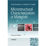
David Brandon, Israel Institute of Technology, Haifa, Israel, is the author of Microstructural Characterization of Materials, 2nd Edition, published by Wiley.
Wayne D. Kaplan, Israel Institute of Technology, Haifa, Israel, is the author of Microstructural Characterization of Materials, 2nd Edition, published by Wiley.
| Preface to the Second Edition | p. xi |
| Preface to the First Edition | p. xiii |
| The Concept of Microstructure | p. 1 |
| Microstructural Features | p. 7 |
| Struture-Property Relationships | p. 7 |
| Microstructural Scale | p. 10 |
| Microstructural Parameters | p. 19 |
| Crystallography and Crystal Structure | p. 24 |
| Interatomic Bonding in Solids | p. 25 |
| Crystalline and Amorphous Phases | p. 30 |
| The Crystal Lattice | p. 30 |
| Summary | p. 46 |
| Bibliography | p. 46 |
| Worked Examples | p. 46 |
| Problems | p. 51 |
| Diffraction Analysis of Crystal Structure | p. 55 |
| Scattering of Radiation by Crystals | p. 56 |
| The Laue Equations and Bragg's Law | p. 56 |
| Allowed and Forbidden Reflections | p. 59 |
| Reciprocal Space | p. 60 |
| The Limiting Sphere Construction | p. 60 |
| Vector Representation of Bragg's Law | p. 61 |
| The Reciprocal Lattice | p. 61 |
| X-Ray Diffraction Methods | p. 63 |
| The X-Ray Diffractometer | p. 67 |
| Powder Diffraction-Particles and Polycrystals | p. 73 |
| Single Crystal Laue Diffraction | p. 76 |
| Rotating Single Crystal Methods | p. 78 |
| Diffraction Analysis | p. 79 |
| Atomic Scattering Factors | p. 80 |
| Scattering by the Unit Cell | p. 81 |
| The Structure Factor in the Complex Plane | p. 83 |
| Interpretation of Diffracted Intensities | p. 84 |
| Errors and Assumptions | p. 85 |
| Electron Diffraction | p. 90 |
| Wave Properties of Electrons | p. 91 |
| Ring Patterns, Spot Patterns and Laue Zones | p. 94 |
| Kikuchi Patterns and Their Interpretation | p. 96 |
| Summary | p. 98 |
| Bibliography | p. 103 |
| Worked Examples | p. 103 |
| Problems | p. 114 |
| Optical Microscopy | p. 123 |
| Geometrical Optics | p. 125 |
| Optical Image Formation | p. 125 |
| Resolution in the Optical Microscope | p. 130 |
| Depth of Field and Depth of Focus | p. 133 |
| Construction of The Microscope | p. 134 |
| Light Sources and Condenser Systems | p. 134 |
| The Specimen Stage | p. 136 |
| Selection of Objective Lenses | p. 136 |
| Image Observation and Recording | p. 139 |
| Specimen Preparation | p. 143 |
| Sampling and Sectioning | p. 143 |
| Mounting and Grinding | p. 144 |
| Polishing and Etching Methods | p. 145 |
| Image Contrast | p. 148 |
| Reflection and Absorption of Light | p. 149 |
| Bright-Field and Dark-Field Image Contrast | p. 150 |
| Confocal Microscopy | p. 152 |
| Interference Contrast and Interference Microscopy | p. 152 |
| Optical Anisotropy and Polarized Light | p. 157 |
| Phase Contrast Microscopy | p. 163 |
| Working with Digital Images | p. 165 |
| Data Collection and The Optical System | p. 165 |
| Data Processing and Analysis | p. 165 |
| Data Storage and Presentation | p. 166 |
| Dynamic Range and Digital Storage | p. 167 |
| Resolution, Contrast and Image Interpretation | p. 170 |
| Summary | p. 171 |
| Bibliography | p. 173 |
| Worked Examples | p. 173 |
| Problems | p. 176 |
| Transmission Electron Microscopy | p. 179 |
| Basic Principles | p. 185 |
| Wave Properties of Electrons | p. 185 |
| Resolution Limitations and Lens Aberrations | p. 187 |
| Comparative Performance of Transmission and Scanning Electron Microscopy | p. 192 |
| Specimen Preparation | p. 194 |
| Mechanical Thinning | p. 195 |
| Electrochemical Thinning | p. 198 |
| Ion Milling | p. 199 |
| Sputter Coating and Carbon Coating | p. 201 |
| Replica Methods | p. 202 |
| The Origin of Contrast | p. 203 |
| Mass-Thickness Contrast | p. 205 |
| Diffraction Contrast and Crystal Lattice Defects | p. 205 |
| Phase Contrast and Lattice Imaging | p. 207 |
| Kinematic Interpretation of Diffraction Contrast | p. 213 |
| Kinematic Theory of Electron Diffraction | p. 213 |
| The Amplitude-Phase Diagram | p. 213 |
| Contrast From Lattice Defects | p. 215 |
| Stacking Faults and Anti-Phase Boundaries | p. 216 |
| Edge and Screw Dislocations | p. 218 |
| Point Dilatations and Coherency Strains | p. 219 |
| Dynamic Diffraction and Absorption Effects | p. 221 |
| Stacking Faults Revisited | p. 227 |
| Quantitative Analysis of Contrast | p. 230 |
| Lattice Imaging at High Resolution | p. 230 |
| The Lattice Image and the Contrast Transfer Function | p. 230 |
| Computer Simulation of Lattice Images | p. 231 |
| Lattice Image Interpretation | p. 232 |
| Scanning Transmission Electron Microscopy | p. 234 |
| Summary | p. 236 |
| Bibliography | p. 238 |
| Worked Examples | p. 238 |
| Problems | p. 247 |
| Scanning Electron Microscopy | p. 261 |
| Components of The Scanning Electron Microscope | p. 262 |
| Electron Beam-Specimen Interactions | p. 264 |
| Beam-Focusing Conditions | p. 265 |
| Inelastic Scattering and Energy Losses | p. 266 |
| Electron Excitation of X-Rays | p. 269 |
| Characteristic X-Ray Images | p. 271 |
| Backscattered Electrons | p. 277 |
| Image Contrast in Backscattered Electron Images | p. 279 |
| Secondary Electron Emission | p. 280 |
| Factors Affecting Secondary Electron Emission | p. 283 |
| Secondary Electron Image Contrast | p. 286 |
| Alternative Imaging Modes | p. 288 |
| Cathodoluminescence | p. 288 |
| Electron Beam Induced Current | p. 288 |
| Orientation Imaging Microscopy | p. 289 |
| Electron Backscattered Diffraction Patterns | p. 289 |
| OIM Resolution and Sensitivity | p. 291 |
| Localized Preferred Orientation and Residual Stress | p. 292 |
| Specimen Preparation and Topology | p. 294 |
| Sputter Coating and Contrast Enhancement | p. 295 |
| Fractography and Failure Analysis | p. 295 |
| Stereoscopic Imaging | p. 298 |
| Parallax Measurements | p. 298 |
| Focused Ion Beam Microscopy | p. 301 |
| Principles of Operation and Microscope Construction | p. 302 |
| Ion Beam-Specimen Interactions | p. 304 |
| Dual-Beam FIB Systems | p. 306 |
| Machining and Deposition | p. 306 |
| TEM Specimen Preparation | p. 310 |
| Serial Sectioning | p. 314 |
| Summary | p. 315 |
| Bibliography | p. 318 |
| Worked Examples | p. 318 |
| Problems | p. 326 |
| Microanalysis in Electron Microscopy | p. 333 |
| X-Ray Microanalysis | p. 334 |
| Excitation of Characteristic X-Rays | p. 334 |
| Detection of Characteristic X-Rays | p. 338 |
| Quantitative Analysis of Composition | p. 343 |
| Electron Energy Loss Spectroscopy | p. 357 |
| The Electron Energy-Loss Spectrum | p. 360 |
| Limits of Detection and Resolution in EELS | p. 361 |
| Quantitative Electron Energy Loss Analysis | p. 364 |
| Near-Edge Fine Structure Information | p. 365 |
| Far-Edge Fine Structure Information | p. 366 |
| Energy-Filtered Transmission Electron Microscopy | p. 367 |
| Summary | p. 370 |
| Bibliography | p. 375 |
| Worked Examples | p. 375 |
| Problems | p. 386 |
| Scanning Probe Microscopy and Related Techniques | p. 391 |
| Surface Forces and Surface Morphology | p. 392 |
| Surface Forces and Their Origin | p. 392 |
| Surface Force Measurements | p. 396 |
| Surface Morphology: Atomic and Lattice Resolution | p. 397 |
| Scanning Probe Microscopes | p. 400 |
| Atomic Force Microscopy | p. 403 |
| Scanning Tunnelling Microscopy | p. 410 |
| Field-Ion Microscopy and Atom Probe Tomography | p. 413 |
| Identifying Atoms by Field Evaporation | p. 414 |
| The Atom Probe and Atom Probe Tomography | p. 416 |
| Summary | p. 417 |
| Bibliography | p. 420 |
| Problems | p. 420 |
| Chemical Analysis of Surface Composition | p. 423 |
| X-Ray Photoelectron Spectroscopy | p. 424 |
| Depth Discrimination | p. 426 |
| Chemical Binding States | p. 428 |
| Instrumental Requirements | p. 429 |
| Applications | p. 431 |
| Auger Electron Spectroscopy | p. 431 |
| Spatial Resolution and Depth Discrimination | p. 433 |
| Recording and Presentation of Spectra | p. 434 |
| Identification of Chemical Binding States | p. 435 |
| Quantitative Auger Analysis | p. 436 |
| Depth Profiling | p. 437 |
| Auger Imaging | p. 438 |
| Secondary-Ion Mass Spectrometry | p. 440 |
| Sensitivity and Resolution | p. 442 |
| Calibration and Quantitative Analysis | p. 444 |
| SIMS Imaging | p. 445 |
| Summary | p. 446 |
| Bibliography | p. 448 |
| Worked Examples | p. 448 |
| Problems | p. 453 |
| Quantitative and Tomographic Analysis of Microstructure | p. 457 |
| Basic Stereological Concepts | p. 458 |
| Isotropy and Anisotropy | p. 459 |
| Homogeneity and Inhomogeneity | p. 461 |
| Sampling and Sectioning | p. 463 |
| Statistics and Probability | p. 466 |
| Accessible and Inaccessible Parameters | p. 467 |
| Accessible Parameters | p. 468 |
| Inaccessible Parameters | p. 476 |
| Optimizing Accuracy | p. 481 |
| Sample Size and Counting Time | p. 483 |
| Resolution and Detection Errors | p. 485 |
| Sample Thickness Corrections | p. 487 |
| Observer Bias | p. 489 |
| Dislocation Density Revisited | p. 490 |
| Automated Image Analysis | p. 491 |
| Digital Image Recording | p. 494 |
| Statistical Significance and Microstructural Relevance | p. 495 |
| Tomography and Three-Dimensional Reconstruction | p. 495 |
| Presentation of Tomographic Data | p. 496 |
| Methods of Serial Sectioning | p. 498 |
| Three-Dimensional Reconstruction | p. 499 |
| Summary | p. 500 |
| Bibliography | p. 503 |
| Worked Examples | p. 503 |
| Problems | p. 514 |
| Appendices | p. 517 |
| Useful Equations | p. 517 |
| Interplanar Spacings | p. 517 |
| Unit Cell Volumes | p. 518 |
| Interplanar Angles | p. 518 |
| Direction Perpendicular to a Crystal Plane | p. 519 |
| Hexagonal Unit Cells | p. 520 |
| The Zone Axis of Two Planes in the Hexagonal System | p. 521 |
| Wavelengths | p. 521 |
| Relativistic Electron Wavelengths | p. 521 |
| X-Ray Wavelengths for Typical X-Ray Sources | p. 521 |
| Index | p. 523 |
| Table of Contents provided by Ingram. All Rights Reserved. |
The New copy of this book will include any supplemental materials advertised. Please check the title of the book to determine if it should include any access cards, study guides, lab manuals, CDs, etc.
The Used, Rental and eBook copies of this book are not guaranteed to include any supplemental materials. Typically, only the book itself is included. This is true even if the title states it includes any access cards, study guides, lab manuals, CDs, etc.