did-you-know? rent-now

Amazon no longer offers textbook rentals. We do!

did-you-know? rent-now

Amazon no longer offers textbook rentals. We do!

We're the #1 textbook rental company. Let us show you why.

9783877065488

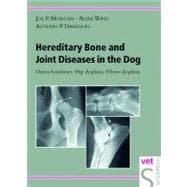
Hereditary Bone and Joint Diseases in the Dog Osteochondroses, Hip dysplasia, Elbow dysplasia

by ; ;
  • ISBN13:

    9783877065488

  • ISBN10:

    3877065481

  • Format: Hardcover
  • Copyright: 2001-04-30
  • Publisher: Schlütersche

Note: Supplemental materials are not guaranteed with Rental or Used book purchases.

Purchase Benefits

List Price: $87.46 Save up to $32.36
  • Rent Book $55.10
    Add to Cart Free Shipping Icon Free Shipping

    TERM
    PRICE
    DUE
    USUALLY SHIPS IN 3-5 BUSINESS DAYS
    *This item is part of an exclusive publisher rental program and requires an additional convenience fee. This fee will be reflected in the shopping cart.

Supplemental Materials

What is included with this book?

Summary

Announcing an unprecedented new source of unique content that goes far beyond a description of bone and joint disorders alone. Every chapter provides information on the history, pathogenesis, diagnosis (physical and radiographic), therapy and prognosis of a particular canine skeletal diseases as well as what the diseases will mean to the dog's life. Expert authors with years of professional experience detail basic and advanced information. Text covers all clinically relevant physical regions in the dog, eg, shoulder, elbow, hip, stifle and tarsus, and presents a group of actual, clinical osteochondrosis cases involving different anatomical locations. This book addresses the importance of selection of dogs for breeding, including changes in breed appearance and disease predilection and the effect of high-energy diets in fast-growing animals. Discussion includes the impact of a lifetime of pain for the affected dog and the treatment expense for clients, yet provides steps that help the owner curb the progressive aspects of bone or joint diseases and manage the anima's discomfort. As a small animal clinician, client questions about skeletal disorders are routine in your practice day. This remarkable, instructional text will give your answers, incidence figures, advice about surgery and timing , and honest analyses of treatment failures and successes. Here is a fresh look at the way we treat OCD, dysplasiias and other bone diseases. With realistic assessments and positives directives for pet care and client support.

Table of Contents

Introduction
Normal growth and ossification of the skeleton
3(1)
Definition of osteochondrosis (OC) and osteochondritis dissecans (OCD)
4(1)
Epiphyseal growth plate
5(3)
Metaphyseal growth plate
8(1)
Definition of dysplasia
9(1)
Definition of osteoarthrosis (OA)
10(2)
Influence of diet and growth rate
12(2)
Heritability of hip dysplasia, elbow dysplasia, and osteochondrosis in the dog
14(1)
Sites of disease predilection in the dog
15(1)
Diagnosis
16(1)
Treatment and prognosis
16(1)
Significance of osteochondrosis and dysplasia
16(1)
Goals of the monograph
17(1)
References
18(3)
Osteochondrosis of the humeral head
Introduction
21(1)
History
21(1)
Course of the disease
22(1)
Gross lesion
23(1)
Clinical signs
24(2)
Physical examination
26(1)
Anatomy and development of the shoulder joint
27(1)
Radiographic diagnosis
27(2)
Radiographic grading of lesions
29(5)
Differential radiographic diagnosis
34(1)
Treatment
35(1)
Conservative treatment
35(1)
Surgical treatment
36(1)
Prognosis
37(1)
Heritability
37(2)
References
39(2)
Elbow dysplasia
Introduction
41(1)
History
41(7)
Proposed etiologies
46(2)
Clinical signs
48(3)
Physical examination
51(1)
Anatomy and development of the elbow joint
51(3)
Radiographic diagnosis
54(3)
Radiographic views
54(2)
Purpose of study
56(1)
Techniques for evaluation
57(1)
Radiographic appearance of the normal elbow joint
57(1)
Radiographic appearance of the dysplastic elbow joint
58(3)
Differential radiographic diagnosis
61(2)
Treatment
63(4)
Conservative treatment
65(1)
Surgical treatment
65(2)
Prognosis
67(1)
Non-operated dogs
67(1)
Operated dogs
67(1)
Immature dogs
67(1)
Mature dogs
68(1)
Heritability
68(1)
Ununited anconeal process (UAP)
69(1)
Introduction
69(2)
History
71(2)
Clinical signs
73(1)
Physical examination
74(1)
Radiographic diagnosis
74(2)
Differential radiographic diagnosis
76(1)
Treatment
76(1)
Conservative treatment
76(1)
Surgical treatment
76(1)
Prognosis
77(1)
Heritability
77(1)
Medial coronoid process disease (MCPD)
77(1)
Introduction
77(1)
History
77(2)
Clinical signs
79(1)
Physical examination
80(1)
Radiographic diagnosis
80(4)
Diagnosis using other radiographic imaging techniques
83(1)
Differential radiographic diagnosis
84(1)
Treatment
85(1)
Conservative treatment
85(1)
Surgical treatment
85(1)
Prognosis
85(1)
Heritability
86(1)
Osteochondrosis of the medial aspect of the humeral condyle
87(1)
Introduction
87(1)
History
87(1)
Clinical signs
88(1)
Physical examination
88(1)
Radiographic diagnosis
89(1)
Differential radiographic diagnosis
89(1)
Treatment
90(1)
Prognosis
90(1)
Heritability
90(1)
References
91(4)
Retained cartilage core
Introduction
95(1)
History
95(1)
Course of the disease
96(1)
Clinical signs
97(1)
Physical examination
97(1)
Anatomy and development of the distal ulna
97(1)
Radiographic diagnosis
98(8)
Differential radiographic diagnosis
106(1)
Treatment
106(1)
Conservative treatment
106(1)
Surgical treatment
106(1)
Prognosis
107(1)
Heritability
107(1)
References
107(2)
Hip dysplasia
Introduction
109(1)
History
109(3)
Affected breeds
112(1)
Prevalence of hip dysplasia
112(1)
Clinical signs
113(5)
Clinical signs in the puppy
113(2)
Clinical signs in the preadolescent
115(1)
Clinical signs in the young dog
116(1)
Clinical signs in the adult dog
117(1)
Clinical signs of lumbosacral disease
117(1)
Clinical signs of transitional lumbosacral vertebral segment
118(1)
Physical examination
118(13)
Joint laxity as an indicator of hip dysplasia
118(2)
Bardens' technique
120(1)
Ortolani technique
121(4)
Anatomy and development of the hip joint
125(1)
Femoral head
126(1)
Acetabulum
126(2)
Hip joint
128(2)
Femoral neck
130(1)
Greater trochanter
131(1)
Radiographic views
131(12)
Ventrodorsal view
133(3)
Ventrodorsal ``frog leg'' view
136(2)
Ventrodorsal wedge view
138(1)
Standing position
139(1)
Badertscher view
140(1)
Fluckiger view
140(1)
Compression-distraction views
141(2)
Dorsal acetabular rim view
143(1)
Radiographic diagnosis
143(25)
Patterns of radiographic change
146(1)
Joint laxity
147(2)
Secondary bony changes
149(5)
Subchondral sclerosis
154(1)
Bilabiation
155(1)
Modeling patterns
156(1)
Acetabular margin
156(1)
Ilial and ischial new bone
156(1)
Femoral head and neck
157(1)
Inclination of the femoral neck
158(4)
Anteversion/retroversion
162(1)
Greater trochanter
163(2)
Pelvic conformation
165(3)
Grading radiographic changes
168(3)
Systems of grading
171(1)
Breed influence on radiographic changes
171(1)
Influence of age on diagnosis
171(4)
Differential radiographic diagnosis
175(9)
Hip luxation
176(1)
Aseptic necrosis of the femoral head
176(3)
Acute fracture
179(1)
Healed fracture of the femoral head or acetabulum
180(1)
Infectious arthritis in the older dog
180(3)
Primary arthrosis
183(1)
Treatment
184(13)
Conservative treatment in the immature dog
184(1)
Conservative treatment in the mature dog
184(1)
Medical treatment
185(1)
Surgical treatment
186(1)
Surgical treatment in the immature dog
187(1)
Tension relief procedures
188(1)
Triple pelvic osteotomy
189(3)
Intertrochanteric derotational varus osteotomy
192(1)
Neck lengthening or intertrochanteric lateralization of the proximal femur
193(1)
Greater trochanteric repositioning
193(1)
Acetabuloplasty
193(1)
Surgical treatment in the mature dog
194(1)
Femoral head and neck ostectomy
194(2)
Total hip replacement
196(1)
Prognosis
197(1)
Heritability
197(6)
References
203(6)
Lumbosacral disease
Introduction
209(1)
History
210(2)
Clinical signs
212(2)
Physical examination
214(1)
Anatomy and development of the lumbosacral region
215(1)
Radiographic diagnosis
216(4)
Survey radiography
216(1)
Stress radiography
216(1)
Epidurography
217(1)
Discography
218(1)
Myelography
218(2)
Examination protocol
220(1)
Differential radiographic diagnosis
220(1)
Treatment
220(3)
Medical treatment
220(1)
Chemonucleolysis
221(1)
Surgical treatment
221(2)
Prognosis
223(1)
Transitional lumbosacral vertebrae
223(1)
Definition
223(1)
Radiographic diagnosis
223(3)
Heritability
226(1)
Osteochondrosis of the sacrum
227
Definition
226(1)
Radiographic diagnosis
227(1)
Heritability
227(1)
Spinal canal stenosis
227(1)
Definition
227(1)
Radiographic diagnosis
228(1)
Heritability
228(1)
References
229(2)
Osteochondrosis of the femoral condyle
Introduction
231(1)
Clinical signs
231(1)
Physical examination
232(1)
Anatomy and development of the femoral condyles
233(1)
Radiographic diagnosis
233(3)
Differential radiographic diagnosis
236(1)
Treatment
236(1)
Conservative treatment
236(1)
Surgical treatment
237(1)
Prognosis
237(1)
Heritability
237(1)
References
238(1)
Osteochondrosis of the talus
Introduction
239(1)
Clinical signs
239(1)
Physical examination
239(1)
Anatomy and development of the talus
240(1)
Radiographic diagnosis
241(2)
Differential radiographic diagnosis
243(1)
Treatment
244(1)
Conservative treatment
244(1)
Surgical treatment
244(1)
Prognosis
244(1)
Heritability
244(1)
References
245(2)
Bone dysplasias in the Labrador Retriever: A radiographic study
Introduction
247(1)
Materials and methods
247(1)
Radiographic studies
247(1)
Results
248(2)
Discussion
250(1)
Conclusions
251(1)
References
252(1)
Schemes for control of disease
Introduction to control of dysplasia through use of registries
253(2)
Control of hip dysplasia in Sweden
255(8)
Joe P. Morgan
Lars Audell
Federation Cynologigue Internationale (FCI)
263(4)
Saki Paatsama
Hip dysplasia and its control in Great Britain
267(17)
Donald D. Lawson
Orthopedic Foundation for Animals, Inc.
284(5)
E.A. Corley
Control of CHD in Switzerland
289(6)
Mark Fluckiger
The Institute for Genetic Disease Control in Animals (GDC)
295(6)
P.W. Poulos
Hip dysplasia diagnosis-the use of distraction radiography
301(4)
G. K. Smith
Control of elbow dysplasia in Sweden
305(4)
Lars Audell
International Elbow Working Group (IEWG)
309(4)
Mark Fluckiger
Index 313

Supplemental Materials

What is included with this book?

The New copy of this book will include any supplemental materials advertised. Please check the title of the book to determine if it should include any access cards, study guides, lab manuals, CDs, etc.

The Used, Rental and eBook copies of this book are not guaranteed to include any supplemental materials. Typically, only the book itself is included. This is true even if the title states it includes any access cards, study guides, lab manuals, CDs, etc.

Rewards Program