did-you-know? rent-now

Amazon no longer offers textbook rentals. We do!

did-you-know? rent-now

Amazon no longer offers textbook rentals. We do!

We're the #1 textbook rental company. Let us show you why.

9783540278764

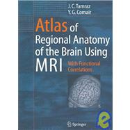
Atlas of Regional Anatomy of the Brain Using MRI : With Functional Correlations

by
  • ISBN13:

    9783540278764

  • ISBN10:

    3540278761

  • Edition: 1st
  • Format: Paperback
  • Copyright: 2006-01-01
  • Publisher: SPRINGER

Note: Supplemental materials are not guaranteed with Rental or Used book purchases.

Purchase Benefits

List Price: $129.00 Save up to $88.84
  • Rent Book $81.27
    Add to Cart Free Shipping Icon Free Shipping

    TERM
    PRICE
    DUE
    USUALLY SHIPS IN 7-10 BUSINESS DAYS
    *This item is part of an exclusive publisher rental program and requires an additional convenience fee. This fee will be reflected in the shopping cart.

Supplemental Materials

What is included with this book?

Summary

The volume provides a unique review of the essential topographical anatomy of the brain from an MRI perspective, correlating high-quality anatomical plates with the corresponding high-resolution MRI images. The book includes a historical review of brain mapping and an analysis of the essential reference planes used for the study of the human brain. Subsequent chapters provide a detailed review of the sulcal and the gyral anatomy of the human cortex, guiding the reader through an interpretation of the individual brain atlas provided by high-resolution MRI. The relationship between brain structure and function is approached in a topographical fashion with analysis of the necessary imaging methodology and displayed anatomy. The central, perisylvian, mesial temporal and occipital areas receive special attention. Imaging of the core brain structures is included. An extensive coronal atlas concludes the book.

Table of Contents

Historical Review of Cross-Sectional Anatomy of the Brain
1(11)
Cephalic Reference Lines Suitable for Neuroimaging
11(40)
Cranial Reference Lines and Planes
11(2)
Historical Background and Overview
11(1)
The Need for a Consensus
12(1)
Classification of the Cephalic Reference Planes
13(1)
The Choice of a Nomenclature
13(1)
Brain Horizontal Reference Lines and Planes
13(26)
The Bicommissural Reference Plane
14(1)
Biometric Data
15(2)
Anatomic and Imaging Correlations
17(5)
The Delmas and Pertuiset Reference Plane
22(1)
Anatomic and Imaging Correlations
22(1)
Topometric Findings
22(2)
The Neuro-ocular Plane
24(1)
Anatomic and Imaging Correlations
24(3)
Topometric and Biometric Findings
27(1)
The Callosal Plane
28(1)
The Chiasmatico-Commissural Plane
29(1)
Biometric Findings
29(2)
Anatomic and Imaging Correlations
31(4)
Anatomic and Physiologic Reference Planes
35(3)
The ``Plan Vestibulaire Horizontal''
38(1)
The ``Plan des Axes Orbitaires''
38(1)
Brain Vertical Reference Lines and Planes
39(12)
The Anterior Commissure-Mamillary Planes
39(1)
The Commissuro-Mamillary Reference Line
39(1)
The Commissuro-Mamillary Plane
40(1)
The Commissural-Obex Reference Plane
41(1)
Biometric Findings
41(1)
Anatomic and Imaging Correlations
42(6)
References
48(3)
Brain Cortical Mantle, Ventricles and White Matter Core
51(66)
Historical Notes and Landmarks
51(1)
Cytoarchitecture and Brain Mapping
51(50)
Gross Morphology and Fissural Patterns of the Brain
56(1)
Gross Morphology
56(1)
Brain Sulcation: Classifications
57(1)
Sulcal and Gyral Anatomy
57(1)
The Lateral Surface of the Cerebral Hemisphere
57(1)
Lateral Fissure of Sylvius
57(12)
Central Sulcus (Rolando)
69(5)
Inferior Frontal Sulcus
74(1)
Superior Frontal Sulcus
74(1)
Precentral Sulcus
74(1)
The Intraparietal Sulcus
74(4)
Superior Temporal Sulcus
78(1)
Frontomarginal Sulcus
78(1)
Gyri of the Lateral Surface of the Cerebral Hemisphere
78(1)
The Frontal Lobe
78(1)
Inferior Frontal Gyrus
78(1)
Middle Frontal Gyrus
78(2)
Superior Frontal Gyrus
80(1)
Precentral Gyrus
80(1)
The Parietal Lobe
80(1)
Postcentral Gyrus
80(1)
Inferior Parietal Gyri
80(1)
Superior Parietal Gyrus
81(1)
The Temporal Lobe
81(1)
Superior Temporal Gyrus
81(1)
Middle Temporal Gyrus
81(1)
Inferior Temporal Gyrus
81(1)
The Occipital Lobe
81(2)
The Insula of Reil
83(1)
The Mesial Surface of the Cerebral Hemisphere
83(3)
Cingulate Sulcus
86(1)
Parieto-occipital Sulcus
86(1)
Calcarine Sulcus
86(1)
Rostral Sulci
86(1)
Gyri of the Mesial Surface of the Cerebral Hemisphere
86(1)
The Gyrus Rectus
86(1)
The Cingulate Gyrus
87(4)
The Medial Frontal Gyrus
91(1)
The Paracentral Lobule
91(1)
The Precuneus
91(1)
The Cuneus
92(1)
The Lingual Gyrus
93(1)
The Basal Surface of the Cerebral Hemisphere
93(1)
The Frontal Orbital Lobe
93(1)
Olfactory Sulcus and Gyrus Rectus
93(1)
Orbital or Orbitofrontal Sulci and Gyri
93(1)
The Temporal Basal Lobe
93(1)
Collateral Sulcus and Parahippocampal Gyrus
93(3)
Occipitotemporal Sulcus and Fusiform Gyrus
96(1)
White Matter Core and Major Association Tracts
96(5)
Vascular Supply of the Brain
101(16)
The Arterial Supply of the Brain
101(1)
The Internal Carotid Artery
101(1)
The Ophthalmic Artery
102(1)
The Posterior Communicating Artery
102(1)
The Anterior Choroidal Artery
103(1)
The Anterior Cerebral Artery
103(2)
The Middle Cerebral Artery
105(1)
The Arterial Circle of Willis
105(5)
Vascular Supply of the Posterior Fossa
110(1)
The Posterior Inferior Cerebellar Artery
110(1)
The Anterior Inferior Cerebellar Artery
111(1)
The Superior Cerebellar Artery
111(1)
The Cerebral Venous System
111(2)
References
113(4)
Central Region and Motor Cortex
117(22)
Introduction
117(1)
Embryology of the Sensorimotor Cortex
117(1)
Morphology and Imaging
117(12)
The Central Sulcus
117(3)
The Precentral Sulcus
120(1)
The Postcentral Sulcus
121(1)
Topographical and Functional Anatomy and Imaging
121(1)
The Primary Sensorimotor Cortex or Central Cortex
121(1)
The Primary Motor Cortex or Precentral Gyrus
122(3)
The Postcentral Gyrus
125(1)
Motor and Sensory Representation in the Primary Sensorimotor Cortex
125(1)
The Premotor Cortex
126(2)
The Supplementary Motor Area
128(1)
The Pyramidal Tract
129(4)
Vascular Supply of the Sensorimotor Cortex
133(2)
Arterial Supply of the Sensorimotor Cortex
133(1)
Arterial Supply of the Lateral Aspect of the Hemisphere
133(1)
Arterial Supply to the Mesial Aspect of the Hemisphere
134(1)
Venous Drainage of the Sensorimotor Cortex
134(1)
The Lateral Venous Drainage System
134(1)
The Mesial Venous Drainage System
135(1)
Imaging Approaches for the Localization of the Central Sulcus
135(4)
References
136(3)
Perisylvian Cognitive Region
139(22)
The Lateral Fissure and the Perisylvian Opercula
139(1)
The Insula of Reil
139(6)
The Preinsular Area
139(1)
The Insular Lobe
139(3)
The Anatomic Relationships of the Insula
142(2)
Vascular Relationships of the Insula
144(1)
Function of the Insula
145(1)
The Anterior Speech Region
145(5)
Sulcal Anatomy of the Anterior Speech Region
146(1)
The Horizontal Ramus
146(1)
The Vertical Ramus
146(3)
The Inferior Precentral Sulcus
149(1)
Gyral Anatomy of the Anterior Speech Region
149(1)
The Pars Triangularis
149(1)
The Pars Opercularis
149(1)
The Posterior Speech Area
150(6)
Sulcal Anatomy of the Posterior Speech Area
151(1)
The Third Segment of the Lateral Fissure
151(1)
The Inferior Parietal Sulci
152(1)
Posterior Extent of the Parallel Sulcus
152(1)
Gyral Anatomy of the Posterior Speech Area
153(1)
Anatomy of Heschl's Gyrus
153(3)
Anatomy of the Temporal Planum
156(1)
Imaging of the Posterior Speech Area
156(1)
The Cerebral Asymmetries
156(5)
References
158(3)
Limbic Lobe and Mesial Temporal Region
161(24)
The Limbic Lobe or ``Grand Lobe Limbique''
161(2)
Morphology and Topographical Anatomy
161(1)
Imaging of the Limbic Lobe
161(2)
The ``Forniceal Plane''
163(1)
Anatomy of the Mesial Temporal Region
163(22)
The Temporal Polar Cortex
163(3)
The Entorhinal Area
166(1)
The Perirhinal Area
166(2)
The Amygdala
168(1)
Morphology, Topographical Anatomy and Imaging of the Amygdala
168(1)
The Basolateral Group of Nuclei
168(1)
The Corticomedial Group of Nuclei
169(1)
The Central Group of Nuclei
170(1)
Boundaries of the Amygdaloid Bodies
170(1)
Functional and Clinical Considerations
171(1)
Clinical Considerations and Targeting
171(1)
The Hippocampal Formation
172(1)
Embryology
172(1)
The Choroid Plexus, Tela, Taenia, and Lamina Affixa
173(1)
Morphology and Imaging
173(1)
Anatomy of the Hippocampal Formation
174(1)
The Hippocampus Proper
174(1)
The Hippocampal Head
174(2)
The Hippocampal Body
176(1)
The Hippocampal Tail
176(1)
Cytoarchitectonics and Intrinsic Circuitry
177(2)
Vascular Supply of the Mesial Temporal Region
179(1)
Functional and Clinical Considerations
179(2)
Clinical Considerations
181(1)
The Uncus
181(1)
References
181(4)
The Basal Forebrain, Diencephalon and Basal Ganglia
185(42)
Regional Anatomy and Imaging of the Interbrain
185(1)
The Basal Forebrain and Related Structures
186(5)
The Septal Region
187(1)
Morphology and Topographical Anatomy and Imaging
187(1)
Functional and Clinical Considerations
188(2)
The Substantia Innominata
190(1)
Morphology and Topographical Anatomy and Imaging
190(1)
Functional and Clinical Considerations
191(1)
The Thalamus
191(9)
Topographical and Nuclear Organization
193(6)
Functional Aspects and Clinical Considerations
199(1)
The Thalamic Radiations
199(1)
The Vascular Supply to the Thalamus
200(1)
The Epithalamus
200(1)
The Pineal Gland
200(1)
The Habenula
200(1)
The Posterior Commissure
200(1)
The Hypothalamus
201(4)
Cytoarchitecture of the Hypothalamus
203(1)
The Medial Hypothalamic Zone
203(1)
The Lateral Hypothalamic Region
203(1)
Connections of the Hypothalamus
204(1)
Functional Aspects and Clinical Considerations
205(1)
Vascular Supply to the Hypothalamus
205(1)
The Subthalamic Region, Subthalamus, or Ventral Thalamus
205(2)
The Subthalamic Nucleus (Corpus Luysi)
206(1)
Morphology and Topographical Anatomy
206(1)
Functional and Clinical Considerations
206(1)
The Zona Incerta
206(1)
The Prerubral Field
207(1)
Vascular Supply to the Subthalamic Region
207(1)
The Basal Ganglia
207(9)
Morphology and Imaging
207(2)
Topographical Anatomy of the Lenticular Nucleus
209(1)
Topographical Anatomy of the Caudate Nucleus
209(3)
Functional Aspects and Clinical Considerations
212(1)
Functional Neurosurgery
213(1)
Arterial Supply to the Basal Ganglia
214(2)
The Internal Capsule and Corona Radiata
216(11)
Gross Morphology and Imaging
216(1)
Topographical and Functional Anatomy
216(1)
Arterial Supply to the Internal Capsule
217(1)
References
217(3)
Synoptical Atlas of Cross-Sectional Anatomy of the Interbrain Using the Commissural-Obex (PC-OB) Reference Plane
220(1)
From the Genu of Corpus Callosum to the Anterior Commissure
220(2)
From the Anterior Commissure to the PC-OB Reference Plane
222(1)
From the PC-OB Plane to the Splenium of Corpus Callosum
223(2)
Abbreviations
225(2)
The Brainstem and Cerebellum
227(30)
Introduction
227(1)
The Brainstem
227(15)
The Midbrain
228(2)
The Rostral Midbrain: Superior Collicular Level
230(1)
The Caudal Midbrain: Inferior Collicular Level
230(1)
The Midbrain-Diencephalic Junction
231(1)
The Red Nuclei and Substantia Nigra
231(1)
The Red Nucleus: Morphology and Functional Anatomy
231(1)
Functional and Clinical Considerations
232(1)
The Substantia Nigra: Morphology and Functional Anatomy
232(2)
Functional and Clinical Considerations
234(1)
The Cerebral Peduncles or Crus Cerebri
235(1)
The Pons
236(1)
Isthmus Rhombencephali: Upper Pons Level
236(1)
The Pons at the Level of the Trigeminal Nerve Root
237(1)
The Pons at the Level of the Advent of the Cerebellum
237(1)
The Medulla Oblongata
238(1)
The Brainstem Reticular Formation
239(1)
Morphology and Topographical Anatomy
239(1)
Functional and Clinical Considerations
239(1)
The Raphe Nuclei or Median Zone
239(1)
The Medial Reticular Zone
240(1)
The Lateral Reticular Zone
240(1)
Vascular Supply to the Brainstem
240(1)
At the Midbrain Level
240(1)
At the Pontine Level
241(1)
At the Medullary Level
241(1)
The Cerebellum
242(15)
Developmental Anatomy and Phylogenesis
242(1)
Morphology and Topographical Anatomy
243(1)
Structural Organization
243(6)
The Deep Cerebellar Nuclei
249(1)
Functional and Clinical Considerations
249(3)
Vascular Supply to the Cerebellum
252(1)
References
253(4)
Optic Pathway and Striate Cortex
257(42)
Introduction
257(1)
Short History of the Anatomy of the Visual Pathways
257(3)
Elements of Ontogenesis, Phylogenesis and Teratology
260(8)
Ontogenesis of the Visual System
260(1)
Phylogenesis of the Anterior Optic Pathways
261(1)
The Eyes
261(1)
The Optic Nerves
262(1)
The Chiasm
262(1)
The Lateral Geniculate Body
262(1)
The Visual Cortex
262(1)
Approach to Evolution Using Magnetic Resonance Imaging
263(1)
Elements of Teratology: The Spectrum of Cyclopia
264(4)
Morphology and Functional Anatomy and Magnetic Resonance Imaging of the Visual System
268(25)
Cephalic Orientations and Visual Pathway
268(1)
The Neuro-ocular Plane
268(1)
Anatomical Imaging Correlations
268(1)
Oculo-orbital Topometry
268(3)
The Chiasmatico-Commissural Plane
271(1)
Anatomic Correlations
271(2)
The Eyes
273(1)
The Optic Nerves
273(1)
The Intraocular Optic Nerve
273(1)
The Intraorbital Optic Nerve
273(1)
The Intracanalicular Optic Nerve
274(6)
The Intracranial Optic Nerve
280(1)
The Optic Chiasm
280(2)
The Optic Tract and the Lateral Geniculate Body
282(3)
The Geniculocalcarine Tract or Optic Radiation
285(3)
The Striate Visual Cortex (Area 17)
288(1)
The Parastriate (Area 18) and Peristriate (Area 19) Cortex
289(1)
The Superior Colliculi as Related to the Visual System
289(4)
Vascular Supply to the Visual Pathway
293(1)
Magnetic Resonance Approach to Neuro-ophthalmological Disorders
293(6)
The Orbital Region
293(1)
The Chiasmal Region
294(1)
The Optic Radiations and Striate Cortex
294(1)
References
295(4)
Atlas of Cross-Sectional Anatomy of the Brain
299(26)
Atlas of Cross-Sectional Anatomy Using the Commissural-Obex Reference Plane (Figs. 10.1, 10.2)
299(13)
From the Frontal Pole to the Genu of the Corpus Callosum (Figs. 10.3--10.10)
299(3)
From the Genu of the Corpus Callosum to the Anterior Commissure (Figs. 10.11--10.16)
302(1)
From the Anterior Commissure to the Commissural-Obex Reference Plane (Figs. 10.17--10.24)
303(3)
From the Commissural-Obex Plane to the Splenium of the Corpus Callosum (Figs. 10.25--10.32)
306(3)
Posterior to the Splenium Toward the Occipital Pole (Figs. 10.33--10.45)
309(1)
Abbreviations
310(2)
Atlas of Cross-Sectional Anatomy Using MR Oriented According to the Ventricular Reference Plane (Figs. 10.46--10.59)
312(4)
Atlas of Cross-Sectional Anatomy Using the Forniceal Reference Plane (Figs. 10.60--10.89)
316(9)
Subject Index 325

Supplemental Materials

What is included with this book?

The New copy of this book will include any supplemental materials advertised. Please check the title of the book to determine if it should include any access cards, study guides, lab manuals, CDs, etc.

The Used, Rental and eBook copies of this book are not guaranteed to include any supplemental materials. Typically, only the book itself is included. This is true even if the title states it includes any access cards, study guides, lab manuals, CDs, etc.

Rewards Program