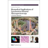
Note: Supplemental materials are not guaranteed with Rental or Used book purchases.
Purchase Benefits
Looking to rent a book? Rent Biomedical Applications of Synchrotron Infrared Microspectroscopy [ISBN: 9780854041541] for the semester, quarter, and short term or search our site for other textbooks by Moss, David. Renting a textbook can save you up to 90% from the cost of buying.
| Fundamentals | |
| Vibrational Spectroscopy: What Does the Clinician Need? | p. 3 |
| Introduction | p. 3 |
| Vibrational Spectroscopy in Cancer | p. 6 |
| Introduction | p. 6 |
| Screening, Early Diagnosis and Surveillance | p. 7 |
| Therapy | p. 9 |
| Vascular Disease | p. 13 |
| Introduction | p. 13 |
| Pathophysiology | p. 13 |
| Vibrational Spectroscopy in Vascular Disease | p. 15 |
| Microbiology and Infective Disease | p. 17 |
| Conclusions | p. 20 |
| References | p. 20 |
| Mid-infrared Spectroscopy: The Basics | p. 29 |
| Introduction | p. 29 |
| Mid-infrared Radiation and Mid-infrared Spectroscopy | p. 30 |
| Electromagnetic Radiation: What is it? | p. 30 |
| Mid-infrared Radiation: What is it? | p. 31 |
| What is Mid-infrared Spectroscopy? | p. 32 |
| Quantitative Mid-infrared Spectroscopy: The Basics | p. 34 |
| Mid-infrared Spectroscopy Instrumentation | p. 35 |
| What is FT-IR? Why FT-IR? | p. 35 |
| FT-IR Microscopy | p. 41 |
| Why Synchrotron-sourced Mid-infrared FT-IR Microspectroscopy? | p. 42 |
| Mid-infrared Spectroscopy: Sampling Techniques and Practices | p. 43 |
| Transmission Sampling Technique | p. 46 |
| Transflection Sampling Technique | p. 47 |
| Attenuated Total Reflection (ATR) Technique | p. 48 |
| ATR Microspectroscopy | p. 51 |
| Near-field FT-IR Microscopy | p. 52 |
| FT-IR Mapping and Imaging Techniques | p. 54 |
| Mid-infrared Spectroscopy: Data Analysis Techniques | p. 55 |
| How Does Mid-infrared Spectroscopy Relate to and Differ from Near-infrared, Far-infrared and Raman Spectroscopy? | p. 56 |
| Fundamental Molecular Vibrations: Mid-infrared and Raman Bands | p. 56 |
| Near-infrared Spectroscopy | p. 61 |
| Far-infrared/THz Spectroscopy | p. 61 |
| Raman Spectroscopy | p. 62 |
| References | p. 63 |
| Infrared Synchrotron Radiation Beamlines: High Brilliance Tools for IR Spectromicroscopy | p. 67 |
| Introduction | p. 67 |
| Infrared Synchrotron Radiation: Historical Background | p. 69 |
| Basic Principles of Synchrotron Radiation | p. 72 |
| Synchrotron Radiation Properties | p. 73 |
| Brilliance | p. 74 |
| Collimation | p. 75 |
| Polarization | p. 76 |
| Stability | p. 76 |
| Time Structure | p. 77 |
| What is an SR Beamline? | p. 78 |
| SR Beamlines and IR Instrumentation for Spectroscopy and Microscopy | p. 82 |
| IR Spectromicroscopy | p. 83 |
| Synchrotron Radiation and Imaging IR | p. 87 |
| Biomedical Applications at IRSR Beamlines | p. 88 |
| Status and Perspectives of IRSR Facilities | p. 94 |
| Conclusions | p. 99 |
| References | p. 100 |
| Raman Microscopy: Complement or Competitor? | p. 105 |
| Introduction | p. 105 |
| Raman Spectroscopy - a Brief History | p. 106 |
| What is Raman Spectroscopy? | p. 108 |
| How is Raman Scattering Measured? | p. 112 |
| System Calibration | p. 118 |
| Raman Spectroscopy for Diagnostics and Biochemical Analysis | p. 122 |
| Raman Microscopy and Imaging at Cellular and Subcellular Levels | p. 127 |
| Comparison to FTIR - Pros and Cons | p. 129 |
| Physical Principles | p. 129 |
| Spatial Resolution | p. 130 |
| Fluorescence and Scattering | p. 133 |
| Photodegradation | p. 134 |
| Signal to Noise | p. 135 |
| Conclusions | p. 137 |
| References | p. 139 |
| Addendum A - Raman Calibration Procedure | p. 142 |
| Technical Aspects | |
| Preparation of Tissues and Cells for Infrared and Raman Spectroscopy and Imaging | p. 147 |
| Introduction | p. 147 |
| Tissue Preparation | p. 148 |
| Introduction to Tissue Preparation Methods | p. 148 |
| Fresh and Cryopreserved Tissue | p. 150 |
| Chemical Fixation of Tissue | p. 152 |
| Paraffin Embedded Tissue | p. 155 |
| Cell Preparation | p. 158 |
| Introduction to Cell Preparation Methods | p. 158 |
| Chemical Fixation of Cells | p. 159 |
| Cell Preparation for Biomechanislic Studies | p. 168 |
| Growth Medium and Substrate Effects on Spectroscopic Examination of Cells | p. 171 |
| Preparation of Living Cells for FTIR and Raman Studies | p. 177 |
| FTIR Studies | p. 177 |
| Raman Studies | p. 180 |
| Conclusions | p. 183 |
| References | p. 185 |
| Data Acquisition and Analysis in Biomedical Vibrational Spectroscopy | p. 192 |
| Introduction | p. 192 |
| Standardisation of the Infrared Spectral Measurements | p. 193 |
| Assessing the Quality of the Obtained Spectra | p. 204 |
| Spectral Pre-processing | p. 206 |
| Data Analysis: Quantitative Analysis | p. 209 |
| Data Analysis: Classification | p. 210 |
| Unsupervised Classification Analysis | p. 210 |
| Supervised Classification Analysis | p. 214 |
| The DPR Approach | p. 216 |
| The Role of Independent Validation | p. 217 |
| Conclusions | p. 220 |
| Acknowledgements | p. 221 |
| Noise and Reproduction Error | p. 221 |
| Differentiation Indices | p. 223 |
| References | p. 223 |
| Synchrotron Radiation as a Source for Infrared Microspec-troscopic Imaging with 2D Multi-Element Detection | p. 226 |
| Introduction | p. 226 |
| Optical Issues for Infrared Microspectroscopy | p. 228 |
| The Standard Infrared Microspectrometer | p. 228 |
| The Schwarzschild Microscope Objective | p. 229 |
| The FPA Infrared Microspectrometer | p. 230 |
| The Synchrotron Infrared Source | p. 232 |
| Basic Properties of the Synchrotron Infrared Source | p. 233 |
| Infrared Microspectroscopy using the Synchrotron Source | p. 234 |
| Imaging at the Diffraction Limit | p. 235 |
| Imaging and the Point Spread Function | p. 235 |
| Performance with the Synchrotron Source and a Single-Element Detector | p. 238 |
| Comparing Synchrotron IR Imaging with Internal Source-based FPA Imaging | p. 240 |
| Diffraction Effects and Issues for PSF Deconvolution | p. 244 |
| Focal Plane Array IR Microspectroscopy with the Synchrotron Source | p. 248 |
| Matching the Dipole Bend Source to the FPA Microspectrometer | p. 249 |
| Initial Results using the Synchrotron Source and FPA | p. 250 |
| Basic PSF Deconvolution with FPA Microspectrometers | p. 253 |
| Opportunities for Advanced 2D Image Deconvolution | p. 254 |
| Conclusions | p. 255 |
| References | p. 256 |
| Scattering in Biomedical Infrared Spectroscopy | p. 260 |
| Introduction to Scattering in Infrared Spectroscopy | p. 260 |
| Mie Scattering | p. 262 |
| Complex Refractive Index | p. 262 |
| The Imaginary Refractive Index, k | p. 262 |
| The Real Refractive Index, n | p. 263 |
| Resonant Mie Scattering (RMieS) | p. 264 |
| Extended Multiplicative Signal Correction (EMSC) | p. 266 |
| Resonant Mie Scattering Correction using the Extended Multiplicative Signal Correction (RMieS-EMSC) | p. 268 |
| Construction of Mie Scattering Efficiency Database | p. 269 |
| Decomposition of the Resonant Mie Scattering Efficiency Database, Q | p. 270 |
| Evaluation of the RMieS-EMSC Algorithm | p. 271 |
| Correction of Real Spectra | p. 272 |
| Conclusions | p. 274 |
| References | p. 275 |
| Case Studies | |
| Synchrotron Based FTIR Spectroscopy in Lung Cancer. Is there a Niche? | p. 279 |
| Introduction | p. 279 |
| Lung Cancer Screening | p. 280 |
| Lung Cancer Diagnosis | p. 283 |
| Treatment of Lung Cancer | p. 286 |
| Conclusions | p. 287 |
| References | p. 287 |
| Head and Neck Cancer: Observations from Synchrotron-sourced Mid-infrared Spectroscopy Investigations | p. 291 |
| Introduction | p. 291 |
| Experimental Work | p. 293 |
| Mid-infrared Synchrotron Radiation FT-IR Studies of Oral Tissue Sections | p. 295 |
| Mid-infrared Synchrotron Radiation FT-IR Studies of Cultured Cells | p. 308 |
| Raman Studies of H&N Samples | p. 312 |
| Conclusions | p. 313 |
| References | p. 314 |
| Single Cell Analysis of TSE-infected Neurons | p. 315 |
| Introduction | p. 315 |
| IR-Spectroscopy and the Composition of Complex Biological Material | p. 316 |
| Why apply Synchrotron FTIR Microspectroscopy (SFTIRM)? | p. 321 |
| Materials and Methods | p. 322 |
| The Study Design | p. 322 |
| Animal Experiments and Sample Preparation | p. 322 |
| Data Acquisition Techniques | p. 323 |
| Data Evaluation Techniques | p. 324 |
| Results | p. 326 |
| Assessment, Discussion and Conclusions | p. 330 |
| Acknowledgements | p. 333 |
| References | p. 333 |
| Monitoring the Effects of Cisplatin Uptake in Rat Glioma Cells: A Preliminary Study Using Fourier Transform Infrared Synchrotron Microspectroscopy | p. 339 |
| Introduction | p. 339 |
| Methodology | p. 341 |
| Cell Culture, Cisplatin Preparation and Treatment | p. 341 |
| Synchrotron FTIR Microspectroscopy | p. 341 |
| Neural Network Classification | p. 342 |
| Results | p. 343 |
| Discussion | p. 346 |
| Conclusions | p. 348 |
| References | p. 349 |
| Mid-Infrared Reflectivity of Mouse Atheromas: A Case Study | p. 351 |
| Introduction | p. 351 |
| Existing Diagnostic Methods | p. 353 |
| Pathologic and Biochemical Features of Vulnerable Plaques | p. 354 |
| Concept of Mid-infrared Reflectivity of Atherosclerotic Aorta | p. 356 |
| Mid-infrared Reflectivity of Experimental Atherosclerosis | p. 358 |
| Discussion | p. 362 |
| Acknowledgements | p. 366 |
| References | p. 366 |
| Subject Index | p. 369 |
| Table of Contents provided by Ingram. All Rights Reserved. |
The New copy of this book will include any supplemental materials advertised. Please check the title of the book to determine if it should include any access cards, study guides, lab manuals, CDs, etc.
The Used, Rental and eBook copies of this book are not guaranteed to include any supplemental materials. Typically, only the book itself is included. This is true even if the title states it includes any access cards, study guides, lab manuals, CDs, etc.