rent-now

Rent More, Save More! Use code: ECRENTAL

5% off 1 book, 7% off 2 books, 10% off 3+ books

9780803611887

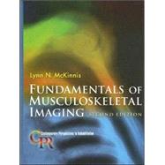
Fundamentals of Musculoskeletal Imaging

by
  • ISBN13:

    9780803611887

  • ISBN10:

    0803611889

  • Edition: 2nd
  • Format: Hardcover
  • Copyright: 2005-06-16
  • Publisher: F a Davis Co
  • Purchase Benefits
  • Free Shipping Icon Free Shipping On Orders Over $35!
    Your order must be $35 or more to qualify for free economy shipping. Bulk sales, PO's, Marketplace items, eBooks and apparel do not qualify for this offer.
  • eCampus.com Logo Get Rewarded for Ordering Your Textbooks! Enroll Now
List Price: $86.00

Summary

A volume in the Contemporary Perspectives In Rehabilitation Series, edited by Steven L. Wolf, PhD, PT, FAPTA.Written specifically for the rehabilitation specialist, this clinically oriented text introduces the basic principles of imaging and the radiographic evaluation of normal and pathologic bone, and fractures. A systems approach organizes chapters by major anatomical region to provide reference points when viewing patient films. IMPORTANT: The answers to the Self-Test Questions in chapters 4 and 6 were inadvertently omitted from the text. To download them, please click the "Online Resources at DavisPlus" link in the "Explore this Product" section at the upper right of this page.

Table of Contents

General Principles of Musculoskeletal Imaging
1(38)
Why Study Imaging?
1(1)
What Is Radiology?
2(1)
What Is Musculoskeletal Imaging?
2(1)
Historical Perspective
2(2)
Turn-of-the-Century Sensationalism
3(1)
1900 to 1930
3(1)
The 1930s
3(1)
The 1940s
3(1)
The 1950s and 1960s
4(1)
The 1970s and 1980s
4(1)
From the 1990s to a New Century
4(1)
Essential Science
4(4)
What Is a Radiograph?
4(1)
What Is Radiation?
5(2)
What Are X-rays?
7(1)
Image Receptors: Different Ways to Capture the X-rays
8(2)
The Gold Standard: Film/Screen Radiography
8(1)
Fluoroscopy
9(1)
Computed Radiography
10(1)
Digital Radiography
10(1)
Understanding the Image
10(6)
What Is Radiodensity?
10(1)
Radiodensity as a Function of Composition: Anatomy in Four Shades of Gray
11(1)
Radiodensity as a Function of Thickness
12(1)
How Many Dimensions Can You See?
12(2)
Radiodensity in a Rose
14(2)
More to the Radiograph
16(10)
Radiographic Terminology
16(1)
Anteroposterior, Lateral, and Oblique Projections
17(3)
Viewing Radiographs
20(1)
Identification Markers
20(1)
Image Quality Factors
21(5)
The Routine Radiographic Examination
26(1)
The Radiologist as the Imaging Specialist
26(2)
Other Common Studies in Musculoskeletal Imaging
28(7)
Contrast-Enhanced Radiographs
28(1)
Conventional Tomography
28(3)
Computed Tomography
31(1)
Nuclear Imaging
31(2)
Magnetic Resonance Imaging
33(1)
Ultrasonography
33(2)
Interventional Techniques
35(1)
Epidural Steroid Injections
35(1)
Spinal Nerve Blocks
35(1)
Radiofrequency Ablation
35(1)
Discography
35(1)
Percutaneous Needle Biopsy of the Spine
36(1)
Percutaneous Vertebroplasty
36(1)
Automated Percutaneous Lumbar Diskectomy
36(1)
Intradiscal Electrothermal Therapy
36(1)
The Imaging Chain
36(1)
Summary of Key Points
37(1)
Self-Test
38(1)
Radiologic Evaluation, Search Patterns, and Diagnosis
39(42)
Where Does Radiologic Image Interpretation Begin?
39(1)
What Are The Pitfalls of Image Interpretation?
39(1)
What Can the Nonradiologist Offer to Image Interpretation?
39(1)
Search Pattern: The ABCs of Radiologic Analysis
40(11)
Alignment
40(3)
Bone Density
43(4)
Cartilage Space
47(2)
Soft Tissues
49(2)
Radiologic Diagnosis of Skeletal Pathology
51(6)
Categories of Skeletal Pathology
52(1)
Distribution of the Lesion
52(1)
Predictor Variables
52(5)
Radiologic Characteristics of Common Pathologies
57(16)
Adult Rheumatoid Arthritis
57(4)
Osteoarthritis (Degenerative Joint Disease)
61(3)
Osteoporosis
64(4)
Musculoskeletal Infections
68(3)
Bone Tumors
71(2)
The Radiological Report
73(5)
Heading
74(1)
Clinical Information
74(2)
Findings
76(1)
Conclusions
76(1)
Recommendations
77(1)
Signature
77(1)
Radiologic Report Example
Summary of Key Points
78(1)
Self-Test
79(2)
Radiologic Evaluation of Fracture
81(36)
Trauma, the Most Common Disorder
81(1)
What Is a Fracture?
82(1)
Biomechanics of Bone
82(1)
Definition of Fracture
82(1)
Trauma Radiology
82(1)
Elements of Fracture Description
83(8)
Anatomic Site and Extent of the Fracture
84(1)
Type of Fracture: Complete or Incomplete
84(1)
Alignment of Fracture Fragments
84(2)
Direction of Fracture Lines
86(2)
Presence of Special Features
88(1)
Associated Abnormalities
88(2)
Fractures Due to Abnormal Stresses or Pathologic Processes
90(1)
Fractures in Children
91(5)
Location Description
91(1)
Difficulties in Assessment of Immature Bone
92(1)
Elements of Fracture Description
92(3)
Healing Factors
95(1)
Reduction and Fixation of Fractures
96(1)
Reduction
96(1)
Fixation
96(1)
Fracture Healing
97(2)
Cortical Bone Healing
97(1)
Cancellous Bone Healing
98(1)
Surgically Compressed Bone Healing
98(1)
Radiologic Evaluation of Healing
98(1)
Time Frame for Fracture Healing
99(2)
Factors That Influence Rate of Fracture Healing
99(2)
Radiologic Examination Intervals During Fracture Healing
101(1)
Complications in Fracture Healing
101(5)
Complications at Fracture Site
101(2)
Late-Effect Complications of Fracture
103(1)
Associated Complications of Other Tissues
104(1)
Life-Threatening Complications
105(1)
Commonly Missed Fractures
106(3)
Clinical History, Evaluation, and Rule of Treatment
106(1)
How Often Are Fractures Missed?
107(2)
Summary of Key Points
109(1)
Case Study
109(2)
Self-Test
111(1)
Appendix: Fracture Eponyms
112(5)
Advanced Imaging: CT, MRI, and Ultrasound
117(24)
Hilmir Agustsson
Computed Tomography
117(5)
History
117(1)
Principles of CT
118(1)
Making the CT Image
118(2)
Viewing CT Images
120(1)
Clinical Uses of CT
121(1)
Summary and Future Developments
122(1)
Magnetic Resonance Imaging
122(8)
History
122(1)
Principles of MRI
123(3)
Making the MR Image
126(1)
Viewing MR Images
127(1)
Clinical Uses of MRI
128(2)
Summary and Future Developments
130(1)
Diagnostic Ultrasound
130(2)
History
130(1)
Principles of Diagnostic Ultrasound
130(2)
Ultrasound Intensity
132(4)
Creating the Ultrasound Image
133(1)
Viewing the Ultrasound Image
133(2)
Clinical Uses of Ultrasound
135(1)
Summary and Future Developments
136(1)
Comparison of Evaluative Properties of CT, MRI, and Ultrasound
136(2)
Summary of Key Points
138(1)
Self-Test
138(3)
Radiologic Evaluation of the Cervical Spine
141(42)
Anatomy Review
141(6)
Osseous Anatomy
141(3)
Ligamentous Anatomy
144(1)
Joint Mobility
145(1)
Growth and Development
146(1)
Postural Development
146(1)
Routine Radiologic Evaluation
147(11)
Practice Guidelines for Spine Radiography in Children and Adults
147(11)
Basic Projections and Radiologic Observations
158
Routine Radiologic Evaluation of the Cervical Spine
148(8)
Optional Projections for Radiologic Evaluation of the Cervical Spine
156(2)
Trauma at the Cervical Spine
158(10)
Radiology for Trauma of the Cervical Spine
158(1)
Potential Injury to Spinal Cord and Spinal Nerves
158(3)
Fractures
161(2)
Dislocations
163(3)
Cervical Spine Sprains
166(1)
Intervertebral Disk Herniations
167(1)
Degenerative Changes in the Cervical Spine
168(3)
Degenerative Disk Disease
169(1)
Degenerative Joint disease
169(1)
Foraminal Encroachment
170(1)
Cervical Spine Spondylosis
170(1)
Spondylosis Deformans
171(1)
Diffuse Idiopathic Skeletal Hyperostosis
171(1)
Clinical Considerations of the Degenerative Spine
171(1)
Cervical Spine Anomalies
171(3)
Summary of Key Points
174(1)
Case Studies
175(5)
Self-Test
180(3)
Radiologic Evaluation of the Temporomandibular Joint
183(26)
Hilmir Agustsson
Historical Perspective
183(1)
The Causes of TMJ Disorders
183(1)
Anatomy Review
184(3)
Osseous Anatomy
184(1)
Ligamentous Anatomy
184(1)
Biomechanics of the TMJ
185(2)
Growth and Development
187(1)
The Role of Imaging in the Evaluation of the TMJ
187(9)
Conventional Radiographs
187(1)
Routine Radiologic Evaluation of the Temporomandibular Joint
188(6)
Conventional Tomography
194(1)
Computed Tomography
194(1)
Magnetic Resonance Imaging
194(2)
Arthritides at the TMJ
196(2)
Osteoarthritis
196(1)
Rheumatoid Arthritis
197(1)
Disk Displacement
198(2)
Etiology
198(1)
Clinical Presentation
198(1)
Classification
198(1)
MR Imaging of Disk Displacements
199(1)
Treatment of Disk Displacement
199(1)
Other Disorders
200(1)
TMJ Hypermobility
200(1)
Fractures
200(1)
Craniomandibular Anomalies
200(1)
The TMJ and the Cervical Spine
201(2)
Positional Faults of the Cervical Spine
202(1)
Acknowledgment
203(1)
Summary of Key Points
204(1)
Case Study
205(1)
Self-Test
206(3)
Thoracic Spine, Sternum, and Ribs
209(40)
Anatomy Review
209(4)
Osseous Anatomy
209(2)
Ligamentous Anatomy
211(1)
Joint Mobility
212(1)
Growth and Development
212(1)
Routine Radiologic Evaluation
213(13)
Practice Guidelines for Spine Radiography in Children and Adults
213(1)
Basic Projections and Radiologic Observations
213(1)
Routine Radiologic Evaluation of the Thoracic Spine
214(4)
Routine Radiologic Evaluation of the Sternum
218(4)
Routine Radiologic Evaluation of the Ribs
222(4)
Trauma at the Thoracic Spine
226(6)
Radiology for Trauma of the Thoracic Spine
226(1)
The Three-Column Concept of Spinal Stability
227(1)
One- or Two-Column Injuries
227(3)
Two- or Three-Column Injuries
230(2)
Fractures of the Bony Thorax
232(1)
Abnormal Conditions
232(8)
Osteoporosis
232(2)
Scoliosis
234(4)
Tuberculous Osteomyelitis (Pott's Disease)
238(1)
Scheuermann's Disease
239(1)
Thoracic, Rib, and Sternal Anomalies
240(2)
Summary of Key Points
242(1)
Case Study
243(4)
Self-Test
247(2)
Lumbosacral Spine and Sacroiliac Joints
249(42)
Anatomy Review
249(6)
Osseous Anatomy
249(2)
Ligamentous Anatomy
251(2)
Joint Mobility
253(1)
Growth and Development
254(1)
Routine Radiologic Evaluation
255(13)
Practice Guideline for Lumbar Spine Radiography in Children and Adults
255(1)
Basic Projections and Radiologic Observations
255(1)
Routine Radiologic Evaluation of the Lumbar Spine
256(8)
Routine Radiologic Evaluation of the Sacroiliac Joint
264(4)
Trauma at the Lumbar Spine
268(5)
Radiology for Trauma of the Lumbar Spine
268(1)
Fractures of the Upper Lumbar Spine
268(1)
Fractures of the Lower Lumbar Spine
268(1)
Spondylolysis
268(2)
Spondylolisthesis
270(3)
Degenerative Conditions at the Lumbar Spine
273(6)
Clinical Considerations of the Degenerative Spine
274(1)
Lumbar Stenosis
274(2)
Intervertebral Disk Herniations
276(3)
Sacroiliac Joint Pathology
279(2)
Ligamentous Injury
279(1)
Degenerative Joint Disease
280(1)
Sacroiliitis
280(1)
Ankylosing Spondylitis
280(1)
Lumbosacral Anomalies
281(3)
Facet Tropism
281(1)
Aberrant Transitional Vertebrae
282(1)
Spina Bifida
283(1)
Summary of Key Points
284(2)
Case Studies
286(4)
Self-Test
290(1)
Radiologic Evaluation of the Pelvis and Hip
291(38)
Anatomy Review
291(3)
Osseous Anatomy
291(2)
Ligamentous Anatomy
293(1)
Joint Mobility
293(1)
Growth and Development
293(1)
Routine Radiologic Evaluation
294(8)
Practice Guidelines for Extremity Radiography in Children and Adults
294(1)
Basic Projections and Radiologic Observations
295(1)
Routine Radiologic Evaluation of the Pelvis
296(2)
Routine Radiologic Evaluation of the Hip and Proximal Femur
298(4)
Trauma at the Pelvis and Hip
302(9)
Radiology for Trauma of the Pelvis and Hip
302(1)
Fractures of the Pelvis
303(3)
Fractures of the Acetabulum
306(2)
Fractures of the Proximal Femur
308(2)
Hip Dislocation
310(1)
Pathological Conditions at the Hip
311(10)
Degenerative Joint Disease of the Hip
311(2)
Rheumatoid Arthritis of the Hip
313(1)
Avascular Necrosis of the Proximal Femur
313(5)
Slipped Femoral Capital Epiphysis
318(1)
Developmental Dysplasia of the Hip
318(3)
Summary of Key Points
321(2)
Case Studies
323(4)
Self-Test
327(2)
Radiologic Evaluation of the Knee
329(38)
Anatomy Review
329(4)
Osseous Anatomy
329(1)
Ligamentous Anatomy
330(1)
Joint Mobility
331(1)
Growth and Development
332(1)
Routine Radiologic Evaluation
333(9)
Practice Guidelines for Knee Radiography in Children and Adults
333(1)
Basic Projections and Radiologic Observations
333(1)
Routine Radiologic Evaluation of the Knee
334(8)
Additional Views Related to the Knee
342(1)
Trauma at the Knee
342(11)
Imaging for Trauma of the Knee
343(1)
Fractures of the Distal Femur
343(1)
Fractures of the Proximal Tibia
344(2)
Fractures of the Patella
346(1)
Subluxations and Dislocations of the Patella
346(1)
Fractures and Dislocations of the Proximal Fibula
347(1)
Fractures of the Articular Cartilage
348(1)
Soft-Tissue Injuries at the Knee
349(4)
Degenerative Joint Disease
353(7)
Radiologic Characteristics
353(2)
Clinical Considerations
355(1)
Treatment
355(1)
Knee Anomalies
355(1)
Genu Valgum
355(1)
Genu Varum
356(1)
Genu Recurvatum
357(3)
Summary of Key Points
360(1)
Case Studies
361(4)
Self-Test
365(2)
Ankle and Foot
367(44)
Anatomy Review
367(21)
Osseous Anatomy
367(1)
Ligamentous Anatomy
368(2)
Joint Mobility
370(1)
Growth and Development
370(18)
Routine Radiologic Evaluation
388(1)
Practice Guidelines for Ankle and Foot Radiography in Children and Adults
388(1)
Basic Projections and Radiologic Observations
388
Routine Radiologic Evaluation of the Ankle
372(10)
Routine Radiologic Evaluation of the Foot
382(6)
Trauma at the Ankle and Foot
388(10)
Radiology for Trauma of the Ankle and Foot
388(1)
Sprains at the Ankle
388(2)
Fractures at the Ankle
390(4)
Fractures of the Foot
394(4)
Deformities of the Foot
398(4)
Radiologic Evaluation
398(1)
Hallux Valgus
398(1)
Pes Cavus
399(1)
Pes Planus
400(1)
Talipes Equinovarus
401(1)
Foot Anomalies
402(1)
Accessory Bones
402(1)
Summary of Key Points
403(1)
Case Studies
404(5)
Self-Test
409(2)
Radiologic Evaluation of the Shoulder
411(36)
Anatomy Review
411(2)
Osseous Anatomy
411(1)
Ligamentous Anatomy
412(1)
Joint Mobility
413(1)
Growth and Development
413(1)
Routine Radiologic Evaluation
413(13)
Practice Guidelines for Shoulder Radiography in Children and Adults
413(13)
Basic Projections and Radiologic Observations
426(12)
Routine Radiologic Evaluation of the Shoulder
416(4)
Routine Radiologic Evaluation of the Acromioclavicular Joint
420(2)
Routine Radiologic Evaluation of the Scapula
422(4)
Additional Views
426(2)
Trauma at the Shoulder
428(1)
Radiology for Trauma of the Shoulder
428(1)
Fractures of the Proximal Humerus
428(2)
Fractures of the Clavicle
430(3)
Fractures of the Scapula
433(1)
Dislocations of the Glenohumeral Joint
434(2)
Acromioclavicular Joint Separation
436(1)
Rotator Cuff Tears
437(1)
Abnormal Conditions at the Shoulder
438(3)
Impingement Syndrome
438(1)
Adhesive Capsulitis
439(2)
Summary of Key Points
441(1)
Case Studies
442(4)
Self-Test
446(1)
Radiologic Evaluation of the Elbow
447(28)
Anatomy Review
447(2)
Osseous Anatomy
447(1)
Ligamentous Anatomy
448(1)
Joint Mobility
448(1)
Growth and Development
448(1)
Routine Radiologic Evaluation
449(13)
Practice Guidelines for Radiography of the Elbow in Children and Adults
449(1)
Basic Projections and Radiologic Observations
449(1)
Routine Radiologic Evaluation of the Elbow
450(8)
Routine Radiologic Evaluation of the Forearm
458(4)
Trauma at the Elbow
462(8)
Radiology for Trauma of the Elbow
462(1)
Fractures of the Distal Humerus
462(3)
Fractures of the Radial Head
465(1)
Fractures of the Proximal Ulna
466(1)
Fractures of the Radial and Ulnar Shafts
467(2)
Dislocations of the Elbow
469(1)
Summary of Key Points
470(1)
Case Studies
470(4)
Self-Test
474(1)
Radiologic Evaluation of the Hand and Wrist
475(56)
Corlia van Rooyen
Anatomy Review
475(4)
Osseous Anatomy
475(1)
Joints and Ligaments of the Hand and Wrist
476(1)
Joint Mobility
477(1)
Growth and Development
478(1)
Routine Radiologic Evaluation
479(19)
Practice Guidelines for Extremity Radiography in Children and Adults
479(19)
Basic Projections and Radiologic Observations
498
Routine Radiologic Evaluation of the Hand
480(6)
Routine Radiologic Evaluation of the Wrist
486(6)
Optional Wrist Views
492(6)
Trauma at the Hand and Wrist
498(11)
General Incidence
498(1)
Radiology for Trauma of the Hand and Wrist
498(1)
Fractures of the Hand
498(4)
Fractures of the Wrist
502(4)
Distal Radius Fractures
506(3)
Wrist Instability
509(10)
Imaging Techniques to Diagnose Instabilty
509(4)
Instability of the Distal Radioulnar Joint
513(2)
Classification of Carpal Instabilities
515(4)
Soft-Tissue Disorders
519(2)
Triangular Fibrocartilage Complex Pathology
519(1)
Carpal Tunnel Syndrome
520(1)
Arthritides
521(2)
Degenerative Joint Disease
521(1)
Rheumatoid Arthritis
522(1)
Summary of Key Points
523(2)
Case Studies
525(5)
Self-Test
530(1)
Integration of Imaging into Physical Therapy Practice
531
J. Bradley Barr
Changing Perspectives on Diagnostic Imaging in Physical Therapy Education
531(3)
The Traditional Model
531(1)
An Evolving Model
531(3)
The Physical Therapist as a Primary Care Provider
534(1)
The Physical Therapist as the Referral Source
534(1)
The Physical Therapist as an Educated User of Diagnostic Imaging
534(1)
The United States Military Health System
535(1)
Other Practice Environments
535(1)
Access to Imaging and Relationships with Physicians
535(2)
The Role of Imaging in the Diagnostic Process
537(1)
When to Recommend Imaging
537(1)
Value of the Information
537(1)
Clinical Decision Making and Clinical Decision Rules
538(3)
The Role of Imaging in Physical Therapy Intervention
541(1)
What Do Physical Therapists Look For?
541(1)
Incorporating Imaging into Treatment Planning
541
Imaging Modalities and Physical Therapy Decision Making
541(1)
What Does the Future Hold?
542(1)
Summary of Key Points
543(1)
Case Studies
544(1)
Self-Test
548

Supplemental Materials

What is included with this book?

The New copy of this book will include any supplemental materials advertised. Please check the title of the book to determine if it should include any access cards, study guides, lab manuals, CDs, etc.

The Used, Rental and eBook copies of this book are not guaranteed to include any supplemental materials. Typically, only the book itself is included. This is true even if the title states it includes any access cards, study guides, lab manuals, CDs, etc.

Rewards Program