rent-now

Rent More, Save More! Use code: ECRENTAL

5% off 1 book, 7% off 2 books, 10% off 3+ books

9780471237662

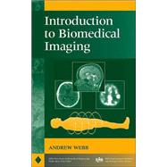
Introduction to Biomedical Imaging

by
  • ISBN13:

    9780471237662

  • ISBN10:

    0471237663

  • Edition: 1st
  • Format: Hardcover
  • Copyright: 2002-12-26
  • Publisher: Wiley-IEEE Press

Note: Supplemental materials are not guaranteed with Rental or Used book purchases.

Purchase Benefits

  • Free Shipping Icon Free Shipping On Orders Over $35!
    Your order must be $35 or more to qualify for free economy shipping. Bulk sales, PO's, Marketplace items, eBooks and apparel do not qualify for this offer.
  • eCampus.com Logo Get Rewarded for Ordering Your Textbooks! Enroll Now
List Price: $216.47 Save up to $54.12
  • Buy Used
    $162.35
    Add to Cart Free Shipping Icon Free Shipping

    USUALLY SHIPS IN 2-4 BUSINESS DAYS

Summary

An integrated, comprehensive survey of biomedical imaging modalitiesAn important component of the recent expansion in bioengineering is the area of biomedical imaging. This book provides in-depth coverage of the field of biomedical imaging, with particular attention to an engineering viewpoint.Suitable as both a professional reference and as a text for a one-semester course for biomedical engineers or medical technology students, Introduction to Biomedical Imaging covers the fundamentals and applications of four primary medical imaging techniques: magnetic resonance imaging, ultrasound, nuclear medicine, and X-ray/computed tomography.Taking an accessible approach that includes any necessary mathematics and transform methods, this book provides rigorous discussions of: The physical principles, instrumental design, data acquisition strategies, image reconstruction techniques, and clinical applications of each modality Recent developments such as multi-slice spiral computed tomography, harmonic and sub-harmonic ultrasonic imaging, multi-slice PET scanning, and functional magnetic resonance imaging General image characteristics such as spatial resolution and signal-to-noise, common to all of the imaging modalities

Author Biography

ANDREW WEBB, PhD, is a faculty member in the Department of Electrical and Computer Engineering and the Beckman Institute for Advanced Science and Technology at the University of Illinois at Urbana-Champaign. Dr. Webb has contributed to many areas of magnetic resonance imaging including developments in radiofrequency coil design, feedback control of thermal processes, techniques for localized spectroscopy, and functional brain mapping. He was awarded a Whitaker Foundation Research Award and a National Science Foundation Career Award in 1997, a Wolfgang-Paul Prize from the Alexander von Humbolt Foundation in 2001, and Xerox and Willett awards for young faculty in 2002. He is a Senior Member of the IEEE.

Table of Contents

Preface.
Acknowledgments.
1. X-Ray Imaging and Computed Tomography.
1.1 General Principles of Imaging with X-Rays.
1.2 X-Ray Production.
1.3 Interactions of X-Rays with Tissue.
1.4 Linear and Mass Attenuation Coefficients of X-Rays in Tissue.
1.5 Instrumentation for Planar X-Ray Imaging.
1.6 X-Ray Image Characteristics.
1.7 X-Ray Contrast Agents.
1.8 X-Ray Imaging Methods.
1.9 Clinical Applications of X-Ray Imaging.
1.10 Computed Tomography.
1.11 Image Processing for Computed Tomography.
1.12 Spiral/Helical Computed Tomography.
1.13 Multislice Spiral Computed Tomography.
1.14 Radiation Dose.
1.15 Clinical Applications of Computed Tomography.
2. Nuclear Medicine.
2.1 General Principles of Nuclear Medicine.
2.2 Radioactivity.
2.3 The Production of Radionuclides.
2.4 Types of Radioactive Decay.
2.5 The Technetium Generator.
2.6 The Biodistribution of Technetium-Based Agents within the Body.
2.7 Instrumentation: The Gamma Camera.
2.8 Image Characteristics.
2.9 Single Photon Emission Computed Tomography.
2.10 Clinical Applications of Nuclear Medicine.
2.11 Positron Emission Tomography.
3. Ultrasonic Imaging.
3.1 General Principles of Ultrasonic Imaging.
3.2 Wave Propagation and Characteristic Acoustic Impedance.
3.3 Wave Reflection and Refraction.
3.4 Energy Loss Mechanisms in Tissue.
3.5 Instrumentation.
3.6 Diagnostic Scanning Modes.
3.7 Artifacts in Ultrasonic Imaging.
3.8 Image Characteristics.
3.9 Compound Imaging.
3.10 Blood Velocity Measurements Using Ultrasound.
3.11 Ultrasound Contrast Agents, Harmonic Imaging, and Pulse Inversion Techniques.
3.12 Safety and Bioeffects in Ultrasonic Imaging.
3.13 Clinical Applications of Ultrasound.
4. Magnetic Resonance Imaging.
4.1 General Principles of Magnetic Resonance Imaging.
4.2 Nuclear Magnetism.
4.3 Magnetic Resonance Imaging.
4.4 Instrumentation.
4.5 Imaging Sequences.
4.6 Image Characteristics.
4.7 MRI Contrast Agents.
4.8 Magnetic Resonance Angiography.
4.9 Diffusion-Weighted Imaging.
4.10 In Vivo Localized Spectroscopy.
4.11 Functional MRI.
4.12 Clinical Applications of MRI.
5. General Image Characteristics.
5.1 Introduction.
5.2 Spatial Resolution.
5.3 Signal-to-Noise Ratio.
5.4 Contrast-to-Noise Ratio.
5.5 Image Filtering.
5.6 The Receiver Operating Curve.
Appendix A: The Fourier Transform.
Appendix B: Backprojection and Filtered Backprojection.
Abbreviations.
Index.

Supplemental Materials

What is included with this book?

The New copy of this book will include any supplemental materials advertised. Please check the title of the book to determine if it should include any access cards, study guides, lab manuals, CDs, etc.

The Used, Rental and eBook copies of this book are not guaranteed to include any supplemental materials. Typically, only the book itself is included. This is true even if the title states it includes any access cards, study guides, lab manuals, CDs, etc.

Rewards Program