did-you-know? rent-now

Amazon no longer offers textbook rentals. We do!

did-you-know? rent-now

Amazon no longer offers textbook rentals. We do!

We're the #1 textbook rental company. Let us show you why.

9781554072378

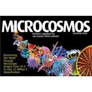
Microcosmos

by
  • ISBN13:

    9781554072378

  • ISBN10:

    1554072379

  • Format: Hardcover
  • Copyright: 2007-09-01
  • Publisher: Firefly Books Ltd
  • Purchase Benefits
  • Free Shipping Icon Free Shipping On Orders Over $35!
    Your order must be $35 or more to qualify for free economy shipping. Bulk sales, PO's, Marketplace items, eBooks and apparel do not qualify for this offer.
  • eCampus.com Logo Get Rewarded for Ordering Your Textbooks! Enroll Now
List Price: $29.95
We're Sorry.
No Options Available at This Time.

Summary

This substantial volume brings together extraordinary images produced through the very latest techniques in microphotography. Most of the 205 stunning, full colour photographs have been taken using scanning electron microscopy (SEM), allowing us to see our world as never before.

Author Biography

Brandon Broil trained as a research biologist before becoming a journalist specializing in science and medicine. He has published stories on subjects as diverse as brain-mapping and crash-test dummies. His work has appeared in numerous international publications, including Reader's Digest and the Guardian, and he has acted as science consultant or editor on many books.

Brandon Broll trained as a research biologist before becoming a journalist specializing in science and medicine. He has published stories on subjects as diverse as brain-mapping and crash-test dummies. His work has appeared in numerous international publications, including Reader's Digest and the Guardian, and he has acted as science consultant or editor on many books.

Table of Contents

Foreword
Microorganisms
Botanics
Human body
Zoology
Minerals
Technology
Photo credits
Table of Contents provided by Publisher. All Rights Reserved.

Supplemental Materials

What is included with this book?

The New copy of this book will include any supplemental materials advertised. Please check the title of the book to determine if it should include any access cards, study guides, lab manuals, CDs, etc.

The Used, Rental and eBook copies of this book are not guaranteed to include any supplemental materials. Typically, only the book itself is included. This is true even if the title states it includes any access cards, study guides, lab manuals, CDs, etc.

Excerpts

ForewordRevealed on these pages is the triumphant power of the scanning electron microscope (SEM) to explore nature and man-made objects at the microscopic level. Combined with the latest techniques microscopists are using to capture the mood and tone of their subjects, and of image artists who use color in order to bring to life the wonder of the microcosmos that surrounds us and is inside us. Most pictures chosen for this book, unless otherwise described, are taken with SEM's. There is reason for this choice. Other microscopes need the specimen to be sliced thinly, or trapped under glass, before it can be examined, creating a two dimensional picture which appears flat or just a part of the whole. A few examples of these methods are illustrated here such as cell cultures and bird flu virus. But the scanning electron microscope, however, reveals another world entirely, a world familiar to the way we naturally see things, a world with outer surfaces and in three-dimensions. How do SEM's achieve this? Electron microscopes rely on a beam of electrons, rather than light, to illuminate a subject. And because an electron beam is produced in a vacuum the specimen must be dead and specially prepared. In SEM'S, a fine electron beam is scanned across the surface of the specimen, highlighting with precision and detail the complexity of the micro-world: microscopic hairs or pores on leaves, the silicate skeletons that make up tiny plankton, even the intricacies of something as mundane as the weave of waterproof clothing or the coil of a lightbulb filament. This power of the scanning electron microscope to resolve microscopic structures can be used to explore the beauty of what is too small to see with the naked eye, but it enjoys other applications in biology and medicine, technology and other sciences. For biologists, certain microorganisms may only be properly identified by their filigree shells as seen under SEM, pollen grains by their sculpted surfaces. In medicine, a nerve cell, for example, can be better understood by researchers able to trace the fine paths of nerve fibers in three-dimensions. In engineering, imperfections on a compact disc can be analysed. Perhaps not realized by the viewer but implicit in these images is that each one has been artificially colored. From a black and white picture produced by SEM, the skilled artist must combine scientific precision with color to make these images come alive. Science has a way of crossing over into art. Not long ago these artists were hand coloring directly onto black and white SEM prints, or using color gels, or chemical stains. Today, however, in our digital age, technology has introduced powerful computing tools that can not only clean and sharpen an image but with masks and layering give it the subtlety of tones in color and depth of shadow found on these pages. Neither are all images here attempts at real-life color. The colors may help to highlight important structures, making a complicated picture simpler to understand. Other images may try to mimic real-life, but only as we imagine it -- such as in our own bodies. Color is a very subjective concept and may just as well be used to enhance a picture's commercial value. For ease of access depending on how much information the reader requires, every picture has a short title, a longer descriptive one, and an in-depth caption. How many times each image has been magnified under the microscope is represented by an X.What follows is a journey into the microcosmos as found around us and inside us in its amazing diversity.Brandon Broll June 2006

Rewards Program