did-you-know? rent-now

Amazon no longer offers textbook rentals. We do!

did-you-know? rent-now

Amazon no longer offers textbook rentals. We do!

We're the #1 textbook rental company. Let us show you why.

9780199637232

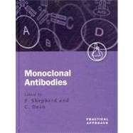
Monoclonal Antibodies A Practical Approach

by ;
  • ISBN13:

    9780199637232

  • ISBN10:

    0199637237

  • Format: Hardcover
  • Copyright: 2000-07-13
  • Publisher: Oxford University Press
  • Purchase Benefits
  • Free Shipping Icon Free Shipping On Orders Over $35!
    Your order must be $35 or more to qualify for free economy shipping. Bulk sales, PO's, Marketplace items, eBooks and apparel do not qualify for this offer.
  • eCampus.com Logo Get Rewarded for Ordering Your Textbooks! Enroll Now
List Price: $234.66
We're Sorry.
No Options Available at This Time.

Summary

Monoclonal Antibodies: A Practical Approach covers the preparation, testing, derivation, and applications of monoclonal antibodies. New immunological techniques incorporating tried and tested methodologies are described, making the book of interest to established and inexperienced immunologists. Both the standard somatic hybridization technique and recombinant techniques, including the use of phage libraries, for the preparation of rodent and human monoclonal antibodies are described. Protocols for both the small and large scale production are detailed, as well as purification and labelling (with both radioisotopes and non-radioisotopes) methods. The applications of monoclonal antibodies in immunoblotting, enzyme linked immunoassays, immunofluorescence, and FACS analysis are all covered in detail. Finally protocols are given for the use of monoclonal antibodies in rheumatoid arthritis, tissue typing, detecting DNA modified during chemotherapy, and in the clinical analysis of transplantation samples for malignancy. This book will therefore be an invaluable laboratory companion to anyone using monoclonal antibodies in their research.

Table of Contents

List of protocols
xix
Abbreviations xxvii
Preparation of rodent monoclonal antibodies by In vitro somatic hybridization
1(24)
Christopher Dean
Philip Shepherd
Introduction and strategy
1(2)
Choice of host for immunization and myeloma for cell fusion
3(1)
Choice of immunogen
4(1)
Preparation of antigen for immunization
5(2)
Soluble antigens
5(1)
Antigens expressed on live cells
5(1)
Plasmid DNA
6(1)
Peptides
7(1)
Route of immunization
7(2)
Generation of immune spleen cells
7(1)
Immunization via the Peyer's patches of rats
7(2)
Growth of myeloma cell lines
9(1)
Preparation of cells for fusion
10(1)
Cell fusion
11(1)
Screening hybridoma culture supernatants for specific antibody
12(6)
Antigen coated multiwell plates
12(4)
Live or fixed cells
16(1)
Ligand binding assays
17(1)
Cloning of hybridomas
18(1)
Characterization and use of the antibodies obtained
19(6)
Isotyping of antibodies
20(1)
Epitope reactivity
20(1)
Specific immunoprecipitation
21(2)
References
23(2)
Preparation of recombinant antibodies from immune rodent spleens and the design of their humanization by CDR grafting
25(33)
Olivier J. P. Leger
Jose W. Saldanha
Introduction
25(1)
Preparation of mouse spleen
26(2)
Isolation of total RNA from spleen
28(2)
Poly(A+) mRNA isolation
30(1)
Reverse transcriptase reaction
31(2)
Primary PCR of antibody genes
33(4)
Preparation of linker
37(2)
Assembly of VH and VK gene fragments with linker DNA
39(2)
Assembly of single chain Fv antibody fragments
41(2)
Reamplification of assembled scFv DNA
43(1)
Restriction enzyme digestion of assembled scFv
44(2)
Purification of pHEN-1 vector by equilibrium centrifugation in CsCI ethidium bromide gradients
46(3)
Restriction digestion of the phage display vector, pHEN-1
49(1)
Ligation of pHEN-1 and insert antibody scFv
50(2)
Preparation of electroporation competent E. coli TG1 strain cells
52(1)
Electroporation
53(2)
Analysis of recombinant clones from the library
55(3)
References
57(1)
Appendix 58(405)
The design of the humanized antibody
58(9)
Issues to consider
59(5)
References
64(3)
Selection of antibodies from phage libraries of immunoglobulin genes
67(24)
Jane K. Osbourn
Introduction
67(1)
Preparation and storage of phage library stocks
68(1)
Phagemid libraries
68(1)
Phage libraries
69(1)
Maintenance of bacterial stocks and titration of phage preparations
69(2)
Selection of phage libraries on purified, immobilized antigen
71(3)
Immobilization of antigen on immunotubes
71(1)
Elution conditions
72(1)
Storage and rescue of the phagemid population after selection
72(1)
Choice of number of selection rounds
73(1)
Selection of phage libraries on biotinylated antigen
74(2)
Biotinylation of antigen
74(1)
Selections using biotinylated antigen
75(1)
Cell surface selections
76(2)
Adherent cell selections
77(1)
Cells in suspension
78(1)
Screening the output of cell surface selections
78(1)
Proximity selections
78(5)
Proximity selection using an existing antibody
80(1)
Proximity selection using natural ligands
81(2)
Step back selections
83(1)
Screening of selected phage
83(4)
Basic screening assays
83(4)
Affinity screening
87(1)
Soluble scFv production and purification
87(4)
References
89(2)
ARM complexes for in vitro display and evolution of antibody combining sites
91(20)
Michael J. Taussig
Maria A. T. Groves
Margit Menges
Hong Liu
Mingyue He
Introduction
91(2)
Ribosome display methodology
93(12)
Outline of procedure
93(1)
Primer design and single chain antibody (VH/K) construction for ARM display
93(3)
Generation of ARM complexes by coupled transcription/translation in vitro
96(2)
Antigen selection of ARM complexes
98(2)
Recovery and amplification of DNA from antigen-selected ARM complexes
100(3)
Further ARM cycles and cloning
103(1)
Analysis of clones encoding antibodies by ARM display
104(1)
Examples of ARM display
105(2)
ARM specificity
105(1)
Selection of DB3R VH/K from libraries
106(1)
Troubleshooting
107(1)
Background
107(1)
No DNA recovery
107(1)
Summary
107(4)
References
109(2)
Human monoclonal antibodies to blood group antigens
111(14)
Belinda M. Kumpel
Introduction
111(1)
General equipment and reagents required
112(1)
Selection of donor
113(1)
Preparation of lymphocytes
113(1)
EBV transformation of B cells
114(3)
Preparation of EBV
115(1)
EBV transformation of B cells and growth of B-LCL
116(1)
Selection of antigen-specific B cells by rosetting
117(1)
Fusion of B-LCL with murine myeloma cells (P3X63Ag8.653)
118(2)
Screening techniques
120(1)
Cloning
121(1)
Cryopreservation of cells
122(3)
References
123(2)
Laboratory based methods for small scale production of monoclonal antibodies
125(24)
Bryan Griffiths
Introduction
125(1)
General principles
125(4)
Culture parameters
125(2)
Medium and serum
127(2)
Stationary cultures
129(2)
Tissue culture plates and flasks
129(1)
Specialized (scale-up) culture systems
130(1)
Stirred cultures
131(4)
Spinner flasks
131(2)
SuperSpinner
133(1)
Stirred bioreactors
134(1)
Dynamic (non-stirred) culture systems
135(2)
Roller bottle culture
135(1)
Airlift fermenters
136(1)
Perfusion (high cell density) systems
137(6)
Hollow fibre (Acusyst) culture
138(1)
Tecnomouse
138(1)
Membrane culture systems (miniPERM)
139(1)
Packed bed systems
140(2)
Fluidized bed bioreactors
142(1)
Harvesting and concentration
143(2)
Harvesting and clarification
143(1)
Concentration
144(1)
Summary
145(4)
References
146(3)
Isolation and purification of monoclonal antibodies from tissue culture supernatant
149(32)
Geoff Hale
Objectives of antibody purification
149(1)
Essential information about the antibody
150(7)
Problems with purifying antibodies from culture supernatant
157
Low concentration of antibody in culture supernatant compared with serum
151(1)
Potential contamination by bovine IgG
152(1)
Equipment for antibody purification
153(3)
Equipment for chromatography
154(2)
Precipitation methods
156(2)
Affinity chromatography
158(4)
Reuse of affinity columns
159(1)
Choice of affinity ligand
160(2)
Immunoaffinity purification
162(1)
Ion exchange chromatography
162(4)
Cation exchange chromatography
163(2)
Anion exchange chromatography
165(1)
Immobilized metal affinity chromatography (IMAC)
166(2)
Size exclusion chromatography (SEC)
168(1)
Hydrophobic interaction chromatography
169(3)
Other chromatographic methods
172(1)
Choice of method
173(1)
Purification of antibody fragments
173(1)
Storage of antibodies
173(1)
Analysis of purity and activity
174(5)
Antibody concentration and purity
174(3)
Endotoxin contamination
177(2)
Conclusion
179(2)
Acknowledgements
179(1)
References
180(1)
Antibody production in plants
181(26)
Pascal Drake
Eva Stoger
Liz Nicholson
Paul Christou
Julian K.-C. Ma
Introduction
181(1)
Expressing recombinant proteins in plants
181(5)
Plant hosts
182(1)
Antibodies in plants
182(3)
Modified viruses for transient expression in plants
185(1)
Glycosylation of recombinant proteins in transgenic plants
185(1)
Plant transformation
186(10)
Gene constructs
187(1)
Agrobacterium tumefaciens-mediated transformation
188(4)
Principles of particle bombardment
192(4)
Plant transformation techniques
196(5)
Agrobaderium-mediated transformation of tobacco
196(3)
Transformation of wheat by micro-projectile bombardment
199(2)
Screening regenerated plantlets for immunoglobulin chain production
201(1)
Self and cross-fertilization of transgenic plants
202(1)
The overall advantages in expressing antibodies in plants
202(5)
References
203(4)
Radiolabelling of monoclonal antibodies
207(30)
Stephen J. Mather
Introduction
207(1)
The choice of radionuclide
207(1)
In vitro applications of radiolabelled antibodies
207(10)
Iodine-125
208(9)
In vivo applications of radiolabelled antibodies
217(17)
Iodine-123
218(1)
Indium-111
219(4)
Technetium-99m
223(8)
Iodine-131
231(1)
Yttrium-90
2(233)
Other therapeutic radionuclides
233(1)
Antibody fragments and genetically engineered constructs
234(1)
Quality control of radiolabelled antibodies
234(3)
References
235(2)
Non-radioactive antibody probes
237(10)
G. Brian Wisdom
Introduction
237(1)
Choice of label
238(1)
General aspects of labelling
238(2)
The labelling reactions
238(1)
The monoclonal antibody
239(1)
Scale and ratios
239(1)
Purification and storage of the labelled antibody
239(1)
Labeiling with an enzyme
240(2)
Labelling with fluorescein
242(1)
Labelling with biotin
243(1)
Labelling with digoxigenin (DIG)
244(1)
Evaluation of labelled monoclonal antibodies
244(3)
References
246(1)
Immunogold probes for light and electron microscopy
247(18)
Paul Monaghan
David Robertson
Introduction
247(2)
Pre-embedding labelling for SEM and TEM
249(2)
Thawed cryosections
251(4)
Progressive lowering of temperature embedding (PLT)
255(1)
Rapid freezing
256(2)
Immunocytochemistry of resin sections
258(3)
Multiple labelling
261(1)
Conclusion
262(3)
References
262(3)
Characterization of cellular antigens using monoclonal antibodies
265(32)
Gillian Hynes
Introduction
265(1)
Initial characterization of monoclonal antibodies
266(1)
Preparation of cell lysates
266(4)
Detection of cellular antigens by immunoblotting
270(13)
Separation of proteins by SDS-PAGE
270(3)
Electrotransfer of proteins from gels to membranes
273(4)
Blocking non-specific binding sites on the membrane
277(1)
Incubation with the primary monoclonal antibody
277(1)
Detection of secondary antibodies and quantitation of signals
277(5)
Troubleshooting problems encountered during immunoblotting
282(1)
Analysis of the electrophoretic properties of polypeptides by 2D PAGE
283(4)
Analysis of subcellular localization of proteins
287(1)
Immunofluorescence staining
287(1)
Subcellular fractionation
287(1)
Resolution of oligomeric complexes by non-denaturing PAGE
287(2)
Detection of cellular antigens by immunoprecipitation
289(6)
Analysis of physiological interactions by immunoprecipitation
292(1)
Immunoprecipitation under disruptive conditions
292(1)
Troubleshooting high background signals
292(3)
Epitope mapping
295(1)
Further applications of monoclonal antibodies in protein characterization
296(1)
Acknowledgements
296(1)
References
296(1)
Immunoassays
297(22)
Jane V. Peppard
Introduction
297(1)
General considerations
297(4)
Selecting an antibody
297(1)
Selecting an assay standard
297(2)
Sample matrix
299(1)
Sample preparation and dilution
299(1)
Assay turnaround time
300(1)
Solid support and separation options
301(2)
Microtitre plates
301(1)
Precipitation from solution
302(1)
Scintillation proximity
303(1)
Labelling and detection of antibody or antigen
303(6)
Enzyme labelling
303(2)
Biotin labelling
305(1)
Labelling with lanthanide fluorophores
306(1)
Radioactive labelling
307(1)
Mass spectrometry
308(1)
Setting up an assay
309(10)
Competitive binding immunoassay
309(2)
'Sandwich' immunoassay
311(4)
Buffers
315(1)
Setting up an ELISA
316(2)
References
318(1)
Immunoaffinity chromatography of macromolecules
319(22)
Steve Hobbs
Introduction
319(1)
Choice of matrix
320(10)
Physical structure
320(1)
Coupling methods
320(1)
Coupling chemistry
321(8)
Monitoring of coupling efficiency
329(1)
Crosslink stability
329(1)
Column storage
330(1)
Sample preparation
330(1)
Binding of antigen
331(3)
Elution
334(1)
Extremes of pH
334(1)
Chaotropic ions
334(1)
Ionic strength
335(1)
Electroelution
335(1)
Antigen reconstitution
335(1)
Analysis of purified antigen
336(1)
Future developments
337(4)
References
338(3)
Immunochemical detection of BrdUrd labelled nuclei for monitoring cell kinetics
341(14)
George D. Wilson
Introduction
341(1)
Basic concepts in cell kinetics
341(2)
Background to the BrdUrd flow cytometry technique
343(2)
In vitro BrdUrd incorporation
345(1)
Fixation procedures
345(1)
Staining procedures
346(3)
DNA denaturation
346(2)
Antibody staining
348(1)
Flow cytometry
349(1)
Examples of data and analysis
350(2)
Conclusions
352(3)
References
352(3)
Immunofluorescence microscopy
355(16)
Rainer Pepperkok
Andreas Girod
Jeremy Simpson
Jens Rietdorf
Introduction
355(1)
Immunofluorescence
355(8)
Fluorescent labelling of antibodies
356(2)
Fixation, permeabilization, and staining of cells
358(5)
Microscopy and imaging equipment
363(5)
Microscopes and associated hardware
363(2)
Imaging detectors for 3D sectioning microscopy
365(2)
Data analysis, image processing, and data presentatio
367(1)
Immunolabelling and visualization in living cells
368(2)
Labelling by microinjection of fluorescent antibodies
368(1)
Microscope set-up for live cell IFM
369(1)
Examples of IFM in fixed cells
370(1)
References
370(1)
FACS analysis of clinical haematological samples in transplantation for cancer
371(20)
Barbara C. Millar
Introduction
371(1)
Fluorescence activated cell analysis
371(6)
General considerations
371(2)
Choice of antibodies, reagents, and general maintenance of the FACS analyser
373(1)
Dual fluorescence
374(2)
Preparation of clinical samples
376(1)
Quality control
376(1)
Calibration of the analyser
377(1)
Assessment of the progenitor cell content in bone marrow (BM) and peripheral blood stem cell (PCSC) harvests
377(6)
Rationale for measuring the progenitor cell content of BM and PBSC harvests for transplantation after intensive therapy for cancer
377(1)
The CD34 determinant
378(1)
Collecting peripheral blood stem cell (PBSC) harvests for CD34+ quantitation
378(1)
Collecting bone marrow (BM) harvests for CD34+ quantitation
379(3)
Quantitation of purified CD34+ cells from PBSC harvests from patients with chronic lymphocytic leukaemia
382(1)
Analysis of lymphocyte subsets in peripheral blood and bone marrow harvests from unrelated donors
383(8)
T cell depletion of paediatric matched (MUD) and unmatched (U-UD) bone marrow (BM) harvests from unrelated donors
383(1)
Analysis of lymphocyte subsets after transplantation or autologous rescue
384(1)
Measurement of intracellular cytokines in mononuclear cells (MNC) and T cells after transplantation or autologous rescue
384(5)
References
389(2)
Immunocytochemical staining of cells and tissues for diagnostic applications
391(20)
Andrew R. Dodson
John P. Sloane
Introduction
391(1)
Tissue and cell substrates
391(1)
Tissue sections
391(1)
Cytological specimens
391(1)
Fixation and processing for paraffin wax embedded tissues
392(1)
Fixation
392(1)
Decalcification
392(1)
Processing
393(1)
Section preparation
393(2)
Section adhesives
393(1)
Section storage
394(1)
Primary antibodies
395(1)
Immunocytochemical methodology
395(7)
Antigen retrieval
396(2)
Detection methodology
398(4)
Reporter molecules
402(3)
Horseradish peroxidase
402(2)
Alkaline phosphatase
404(1)
Enhancement
405(2)
Biotinylated tyramine
406(1)
Controls
407(4)
Positive control
407(1)
Negative control
407(1)
External quality assurance scheme (EQA)
407(1)
Acknowledgements
407(1)
Suggested Further Reading
407(1)
References
408(3)
Detection of chemically modified DNA in lymphocytes of patients undergoing chemotherapy
411(20)
Michael J. Tilby
Introduction
411(3)
Types of modifications to which antibodies can be produced
412(1)
Applications of antibodies against modified DNA
412(1)
Merits of raising antibodies against modified mono/dinucleotides versus modified polymeric DNA
412(2)
Requirements for detection of low frequencies of DNA modifications
414(1)
Production of appropriate antibodies
414(1)
Immunization
414(1)
Screening hybridomas
415(1)
ELISA techniques for quantification of DNA modifications
415(11)
Coating wells with DNA
416(2)
Direct binding assays
418(1)
Competitive assays
419(7)
Staining for adducts in individual cells
426(5)
Staining techniques: conventional versus agarose embedded DNA
426(1)
Staining agarose embedded DNA
427(2)
References
429(2)
Monoclonal antibody therapy in organ transplantation
431(18)
Matt Wise
Diana Zelenika
Introduction
431(1)
The challenge of antibody therapy in humans
432(3)
Immunological tolerance versus immunosuppression
432(1)
Depletion versus non-depletion
433(1)
Avoiding the antiglobulin response
434(1)
Can monoclonal antibodies ever be used to induce tolerance in humans?
434(1)
Animal models of transplantation and mAb therapy: defining the problem
435(1)
Which mAbs for organ transplantation
436(13)
Anti-CD3 mAb treatment
437(3)
Monoclonal antibodies to CD4 and CD8
440(2)
Monoclonal antibodies to CD2S (IL-2 receptor)
442(1)
Blockade of co-stimulation through CD4O and CD28 pathways
443(1)
T cell depletion
444(1)
Summary
445(1)
References
445(4)
Monoclonal antibody therapy In rheumatoid arthritis
449(14)
Ernest H. S. Choy
Gabrielle H. Kingsley
Gabriel S Panayi
Introduction
449(1)
Pathogenesis of rheumatoid arthritis
449(1)
Treatment strategies in rheumatoid arthritis
450(1)
Therapeutic monoclonal antibodies
451(1)
Monoclonal antibodies in rheumatoid arthritis
452(6)
Anti-cytokine mAbs in rheumatoid arthritis
452(2)
Anti-adhesion molecule mAbs
454(1)
Anti-T cell mAbs in RA
455(3)
Conclusion
458(5)
Acknowledgements
459(1)
References
459(4)
List of suppliers 463(6)
Index 469

Supplemental Materials

What is included with this book?

The New copy of this book will include any supplemental materials advertised. Please check the title of the book to determine if it should include any access cards, study guides, lab manuals, CDs, etc.

The Used, Rental and eBook copies of this book are not guaranteed to include any supplemental materials. Typically, only the book itself is included. This is true even if the title states it includes any access cards, study guides, lab manuals, CDs, etc.

Rewards Program