did-you-know? rent-now

Amazon no longer offers textbook rentals. We do!

did-you-know? rent-now

Amazon no longer offers textbook rentals. We do!

We're the #1 textbook rental company. Let us show you why.

9783540255239

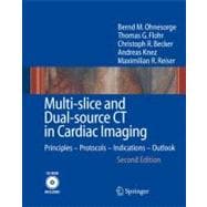
Multi-Slice and Dual-Source CT in Cardiac Imaging: Principles, Protocols, Indications, Outlook

by ; ; ; ;
  • ISBN13:

    9783540255239

  • ISBN10:

    3540255230

  • Edition: 2nd
  • Format: Hardcover
  • Copyright: 2006-12-04
  • Publisher: Springer Verlag

Note: Supplemental materials are not guaranteed with Rental or Used book purchases.

Purchase Benefits

List Price: $219.00 Save up to $148.46
  • Buy Used
    $164.25
    Add to Cart Free Shipping Icon Free Shipping

    USUALLY SHIPS IN 2-4 BUSINESS DAYS

Supplemental Materials

What is included with this book?

Summary

Cardiac diseases, and in particular coronary artery disease, are the leading cause of death and morbidity in industrialized countries. The development of non-invasive imaging techniques for the heart and the coronary arteries has been considered a key element in improving patient care. A breakthrough in cardiac imaging using CT occurred in 1998, with the introduction of multi-slice computed tomography (CT). Since then, amazing advances in performance have taken place with scanners that acquire up to 64 slices per rotation. This book discusses the state-of-the-art developments in multi-slice CT for cardiac imaging as well as those that can be anticipated in the future. It serves as a comprehensive work that covers all aspects of this technology, from the technical fundamentals and image evaluation all the way to clinical indications and protocol recommendations. This fully reworked second edition draws on the most recent clinical experience obtained with 16- and 64-slice CT scanners by world-leading experts from Europe and the United States. It also includes "hands-on" experience in the form of 10 representative clinical case studies, which are included on the accompanying CD. As a further highlight, the latest results of the very recently introduced dual-source CT, which may soon represent the CT technology of choice for cardiac applications, are presented. This book will not only convince the reader that multi-slice cardiac CT has arrived in clinical practice, it will also make a significant contribution to the education of radiologists, cardiologists, technologists, and physicists"whether newcomers, experienced users, or researchers.

Table of Contents

Introduction
1(22)
Bernd Ohnesorge
Basic Principles of CT Imaging
1(2)
Established Imaging Modalities for Cardiac Imaging
3(8)
Invasive Imaging
3(5)
Non-invasive Imaging
8(3)
Clinical Goals for CT in the Diagnosis of Cardiac and Thoracic Diseases
11(3)
Coronary Artery Disease
11(2)
Other Cardiac Diseases
13(1)
Diseases of the Thoracic Vessels
14(1)
The History and Evolution of CT in Cardiac Imaging
14(9)
Principles and Applications of Electron-Beam CT
14(3)
Cardiac Imaging with Conventional Mechanical CT
17(3)
References
20(3)
Cardiac and Cardiothoracic Anatomy in CT
23(18)
Roman Fischbach
Topography
23(1)
Standard Views
24(6)
Coronary Arteries and Veins
30(2)
Pericardium
32(1)
Cardiac Chambers
33(1)
Cardiac Valves
34(1)
Great Vessels
35(6)
References
39(2)
Multi-slice CT Technology
41(30)
Thomas Flohr
Bernd Ohnesorge
Evolution from 1 to 64 Slices
41(4)
Principles of Multi-slice CT System Design
45(7)
Multi-slice CT Acquisition and Reconstruction for Body Imaging
52(19)
Definition of the Pitch
52(1)
The Cone-Angle Problem in Multi-slice CT
53(1)
Multi-slice Spiral Reconstruction Neglecting the Cone-Beam Geometry
54(4)
Multi-slice Spiral Reconstruction with Cone-Beam Algorithms
58(9)
References
67(4)
Principles of Multi-slice Cardiac CT Imaging
71(56)
Bernd Ohnesorge
Thomas Flohr
Basic Performance Requirements for CT Imaging of the Heart
71(2)
CT Imaging with Optimized Temporal Resolution: The Principle of Half-Scan Reconstruction
73(3)
Prospectively ECG-Triggered Multi-slice CT
76(3)
Retrospectively ECG-Gated Multi-slice CT
79(27)
Multi-slice Cardiac Spiral Reconstruction
81(2)
ECG-Gated Multi-slice Spiral Acquisition
83(5)
Segmented Cardiac Reconstruction Algorithms
88(9)
Cardiac Cone-Beam Reconstruction Algorithms
97(4)
ECG-Gated Spiral Scanning with Increased Volume Speed
101(5)
Synchronization with the ECG and Cardiac Motion
106(7)
ECG-Based Phase Selection
106(2)
The Pros and Cons of ECG Gating and ECG Triggering
108(2)
Alternative Cardiac-Motion Gating Approaches
110(3)
Radiation Exposure Considerations
113(14)
Principles of Radiation Dose Measurement in CT
113(4)
Radiation Exposure for Selected Cardiac Examination Protocols
117(2)
Exposure Reduction with ECG-Gated Tube-Current Modulation
119(2)
Optimization for Different Patient Sizes
121(2)
Optimization of Contrast-to-Noise Ratio
123(1)
References
124(3)
Clinical Examination Protocols with 4- to 64-Slice CT
127(24)
Bernd Ohnesorge
Quantification of Coronary Artery Calcification
127(6)
CT Angiography of the Cardiac Anatomy and Coronary Arteries
133(8)
Cardiac Function Imaging
141(3)
Cardiothoracic Examination Protocols
144(7)
References
148(3)
Image Visualization and Post-processing Techniques
151(41)
Thomas Flohr
Bernd Ohnesorge
Trans-axial Image Slices
151(2)
Multi-planar Reformation
153(1)
Maximum-Intensity Projection
154(3)
Volume-Rendering Technique
157(5)
Vessel Segmentation and Vessel Analysis
162(3)
Four-Dimensional Visualization and Functional Parameter Assessment
165(4)
Myocardial Perfusion Evaluation
169(2)
Quantification of Coronary Calcification
171(8)
References
176(3)
Clinical Indications
Current and Future Clinical Potential
179(13)
Christoph Becker
Andreas Knez
Cardiac Multi-slice CT Technique
179(1)
Coronary Calcium Screening
180(2)
Coronary CT Angiography
182(7)
Non-coronary Applications
189(1)
References
190(2)
Risk Assessment with Coronary Artery Calcium Screening
192(13)
Roman Fischbach
Introduction
192(1)
Methods of Coronary Artery Calcification Quantification
192(3)
Multi-slice CT Examination Technique
195(2)
Clinical Applications of Coronary Calcium Measurement
197(3)
Further Clinical Applications of Calcium Scanning
200(2)
Conclusion
202(1)
References
203(2)
Detection and Exclusion of Coronary Artery Stenosis
205
Koen Nieman
N. Mollet
F. Cademartiri
F. de Feyter
Introduction
205(1)
Diagnostic Performance of 4-Slice CT
205(1)
Diagnostic Performance of 16-Slice CT
206(1)
Diagnostic Performance of 64-Slice CT
207(1)
Image Processing and Analysis
208(1)
Discussion
208(4)
References
212
Assessment and Interpretation of Atherosclerotic Coronary Plaques
192(32)
Stephan Achenbach
Clinical Background
214(1)
Clinical Concepts
215(1)
Visualization of Coronary Atherosclerotic Plaques
215(1)
Visualization of Non-calcified Plaques by Multi-slice CT
215(4)
Perspective
219(2)
References
221(3)
Coronary CT Angiography in Patients with Chest Pain
224(8)
Axel Kuttner
Stephen Schroder
Clinical Background
224(1)
Methods and Protocols
225(1)
Results in a Representative Patient Population
226(2)
Discussion
228(2)
References
230(2)
Evaluation of Coronary Artery Bypass Grafts
232(7)
Axel Kuttner
Andreas Kopp
Clinical Background
232(1)
Protocol to Assess Graft Patency and Native Vessels with Multi-slice CT
233(2)
Results in a Representative Patient Population
235(2)
Conclusion
237(1)
References
238(1)
Patency Control of Coronary Stents
239(6)
Koen Nieman
N. Mollet
F. Candemartiry
F. de Feyter
Introduction
239(1)
CT Imaging Characteristics of Stents
239(1)
Post-processing and Analysis
240(1)
In Vivo Coronary Stent Imaging
241(1)
Conclusion
242(2)
References
244(1)
Evaluation of the Coronary Anomaly, Fistula, Aneurysm, and Dissection
245(13)
Steffen Frohner
Introduction
245(1)
Anomalies of the Coronary Arteries
245(1)
Coronary Artery Fistulas
246(5)
Myocardial Bridges
251(3)
Coronary Aneurysms
254(1)
Coronary Dissection
254(2)
Coronary Vasculitis
256(1)
Conclusion
256(1)
References
256(2)
Diagnosis of Congenital Heart Disease in Adults and Children
258(8)
Jean-Francois Paul
Introduction
258(1)
Technical Aspects and Imaging Protocols
258(3)
Clinical Indications
261(2)
Improvements with 64-Slice CT
263(1)
Conclusion
263(2)
References
265(1)
Evaluation of Ventricular Function Parameters
266(15)
Kai Uwe Jurgens
Roman Fischbach
Introduction
266(1)
Determination of Cardiac Function Parameters with Multi-slice CT
267(2)
Data Acquisition and Image Reconstruction
269(1)
Image Analysis
270(1)
Limitations
270(2)
Clinical Considerations
272(5)
Summary and Outlook
277(2)
References
279(2)
Imaging and Diagnosis of Cardiac Valves
281(9)
Jurgen Willmann
Introduction
281(1)
Technical Considerations
281(1)
Valvular Morphology
282(1)
Valvular Disease
283(2)
Limitations
285(1)
Conclusion
286(1)
References
286(4)
Visualization of Cardiac Tumors and Masses
290(6)
Bernd Wintersperger
Introduction
288(2)
Imaging Techniques
290(1)
Epidemiology of Cardiac Masses and Clinical Application of CT
290(3)
Conclusion
293(2)
References
295(1)
Imaging of the Pulmonary Veins in Patients with Atrial Fibrillation
296(11)
Michael Poon
C. Learra
Introduction
296(1)
Pharmacological Treatment Options
297(1)
Non-pharmacological Treatment of Atrial Fibrillation: Catheter Ablation
298(1)
The Role of Imaging in the Era of Catheter Ablation of the Pulmonary Veins
299(1)
Multi-slice CT Imaging of the Pulmonary Veins and Left Atrium
299(3)
The Role of Multi-slice CT in Catheter Ablation of Afib
302(1)
Conclusions
303(2)
References
305(2)
Potential of Myocardial Perfusion and Viability Studies
307(10)
Konstantin Nikolaou
Bernd Wintersperger
Introduction
307(1)
Principles of Myocardial Perfusion Assessment
307(1)
Technical Considerations of Multi-slice CT
308(1)
Study Data on Myocardial Perfusion Imaging Using Multi-slice CT
308(2)
Assessment of Myocardial Viability with Multi-Slice CT
310(3)
Conclusion
313(2)
References
315(2)
Cardiothoracic Multi-Slice CT in the Emergency Department
317(10)
Uwe-Joseph Schopf
ECG-Gated Multi-Slice CT Scanning of the Chest
317(3)
Patients with Equivocal Chest Pain in the Emergency Department
320(1)
64-Slice CT as a Triage Tool in the Emergency Department
320(4)
References
324(3)
Future Technical Developments in Cardiac CT
327(32)
Bernd Ohnesorge
Limitations and Pitfalls with Today's Multi-slice CT
327(5)
Temporal Resolution
327(1)
Volume Coverage
328(1)
Spatial Resolution and Signal-to-Noise Ratio
329(2)
Radiation Exposure
331(1)
A Future for Electron-Beam CT?
332(2)
Future Possibilities with Area Detector CT
334(1)
New Frontiers with Dual-Source CT
335(24)
Dual-Source CT: System Concept and Design
338(1)
Dual-Source CT: Cardiac Scanning Principles and Techniques
339(3)
Dual-Source CT: System Performance Evaluation
342(5)
Dual-Source CT: Clinical Scan Protocols and Preliminary Clinical Results
347(10)
References
357(2)
Subject Index 359

Supplemental Materials

What is included with this book?

The New copy of this book will include any supplemental materials advertised. Please check the title of the book to determine if it should include any access cards, study guides, lab manuals, CDs, etc.

The Used, Rental and eBook copies of this book are not guaranteed to include any supplemental materials. Typically, only the book itself is included. This is true even if the title states it includes any access cards, study guides, lab manuals, CDs, etc.

Rewards Program