did-you-know? rent-now

Amazon no longer offers textbook rentals. We do!

did-you-know? rent-now

Amazon no longer offers textbook rentals. We do!

We're the #1 textbook rental company. Let us show you why.

9783527313839

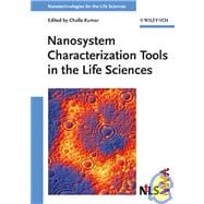
Nanosystem Characterization Tools in the Life Sciences

by
  • ISBN13:

    9783527313839

  • ISBN10:

    3527313834

  • Edition: 1st
  • Format: Hardcover
  • Copyright: 2006-02-10
  • Publisher: Wiley-VCH

Note: Supplemental materials are not guaranteed with Rental or Used book purchases.

Purchase Benefits

  • Free Shipping Icon Free Shipping On Orders Over $35!
    Your order must be $35 or more to qualify for free economy shipping. Bulk sales, PO's, Marketplace items, eBooks and apparel do not qualify for this offer.
  • eCampus.com Logo Get Rewarded for Ordering Your Textbooks! Enroll Now
List Price: $307.19 Save up to $113.66
  • Rent Book $193.53
    Add to Cart Free Shipping Icon Free Shipping

    TERM
    PRICE
    DUE
    USUALLY SHIPS IN 3-4 BUSINESS DAYS
    *This item is part of an exclusive publisher rental program and requires an additional convenience fee. This fee will be reflected in the shopping cart.

Supplemental Materials

What is included with this book?

Summary

This Volume. Volume 3 reviews spectroscopic, scanning and X-ray methodologies for the gleaning of information with nano-scale resolution as well as special characterization techniques and the requirements of the biomedical context of the measurements. Book jacket.

Author Biography

<b>Challa Kumar</b> is currently the Group Leader of Nanofabrication at the Center for Advanced Microstructures and Devices (CAMD), Baton Rouge, USA. His research interests are in developing novel synthetic methods for functional nanomaterials and innovative therapeutic, diagnostic and sensory tools based on nanotechnology. He has eight years of industrial R&amp;D experience working for Imperial Chemical Industries and United Breweries prior to joining CAMD. He is the founding Editor-in-Chief of the Journal of Biomedical Nanotechnology, an international peer reviewed journal published by American Scientific Publishers, and the series editor for the ten-volume book series Nanotechnologies for the Life Sciences (NtLS) published by Wiley-VCH. He worked at the Max Planck Institute for Biochemistry in Munich, Germany, as a post doctoral fellow and at the Max Planck Institute for Carbon Research in Munich, Germany, as an invited scientist. He obtained his Ph.D. degree in synthetic organic chemistry from Sri Sathya Sai Institute of Higher Learning, Prashanti Nilayam, India.

Table of Contents

Preface xiii
List of Contributors xvi
1 Fluorescence Imaging in Biology using Nanoprobes
1(37)
Daniele Gerion
1.1 Introduction and Outlook
1(2)
1.1.1 A New Era in Cell Biology
1(1)
1.1.2 Nanotechnology and its Perspectives for Fluorescence Imaging in Cell Biology
2(1)
1.2 Fundamentals of Fluorescence
3(14)
1.2.1 Basic Principles
3(3)
1.2.2 A Few Types of Fluorescent Probes
6(1)
1.2.2.1 Small Luminescent Units and Autofluorescence of Living Organisms
6(1)
1.2.2.2 A few Organic Dyes and their Limitation in Live Cell Labeling
7(1)
1.2.2.3 Green Fluorescent Protein and its Cousin Mutants
8(1)
1.2.2.4 Quantum Dots
9(1)
1.2.2.5 Toxicity Issues of Nanomaterials
13(1)
1.2.3 Sources and Detectors
14(1)
1.2.3.1 Light Sources
14(1)
1.2.3.2 Detectors
15(2)
1.3 Microscope Configurations
17(4)
1.3.1 Wide-field Methods: Epi-, and Total Internal Reflection (TIR)
17(1)
1.3.1.1 Epifluorescence Illumination
17(1)
1.3.1.2 Total internal Reflection (TIR) Illumination
18(1)
1.3.2 Scanning Methods for Microscopy
19(1)
1.3.2.1 Laser-scanning or Stage-scanning Confocal Microscopy
19(1)
1.3.2.2 Near-field Scanning Optical Microscopy (NSOM)
20(1)
1.4 Strategies for Image Acquisition
21(5)
1.4.1 Intensity Imaging
21(2)
1.4.2 Spectral Imaging
23(1)
1.4.3 Lifetime and Time-gated Imaging
24(2)
1.4.4 Other Imaging Modalities: Polarization and FRET Imaging
26(1)
1.5 Qdots in Biology. A Few Selected Examples
26(5)
1.5.1 Ultra-high Colocalization of Qdots for Genetic Mapping
27(1)
1.5.2 Dynamics of Biomolecules in a Cellular Environment
28(1)
1.5.2.1 Trafficking of Glycine Receptors in Neural Membranes of Live Cells
29(1)
1.5.2.2 Dynamics of Labeled Nuclear Localization Sequences Inside Living Cells
30(1)
1.5.3 In Vivo and Non-invasive Detection Using Qdot Reporters
31(1)
1.6 Outlook: Is there a Role for Nanoscience in Cellular Biology and in Medicine?
31(1)
Acknowledgments
32(1)
References
33(5)
2 Characterization of Nanoscale Systems in Biology using Scanning Probe Microscopy Techniques
38(71)
Anthony W. Coleman, Adina N. Lazar, Cecile F. Rousseau, Sebastien Cecillon, and Patrick Shahgaldian
2.1 Introduction
38(1)
2.2 The Scanning Probe Microscopy Experiment
39(1)
2.3 Scanning Tunneling Microscopy Imaging
40(1)
2.4 Atomic Force Microscopy
41(15)
2.4.1 Generalities
41(3)
2.4.2 Tips and Cantilevers
44(2)
2.4.3 Contact Mode AFM
46(1)
2.4.4 Dynamic Modes
47(1)
2.4.4.1 Generalities
47(1)
2.4.4.2 Non-contact Mode
48(1)
2.4.4.3 Intermittent Contact Mode
49(1)
2.4.4.4 Force Modulation Mode
49(1)
2.4.5 Friction Force Mode or Lateral Force Mode
50(1)
2.4.6 Force–Distance Analysis
50(2)
2.4.7 Chemical Force Imaging
52(2)
2.4.8 Dip-pen Lithography
54(1)
2.4.9 Cantilever Array Sensors
54(2)
2.5 Near-field Scanning Optical Microscopy
56(1)
2.6 Artifacts
57(3)
2.6.1 Artifacts Related to Tip Size and Geometry
57(2)
2.6.2 Artifacts from Damaged Tips
59(1)
2.6.3 Artifacts from Tip–Sample Interactions
59(1)
2.6.4 Sample Artifacts
59(1)
2.7 Using the Tools
60(33)
2.7.1 DNA
60(1)
2.7.1.1 Topographic Imaging of DNA
60(1)
2.7.1.2 Imaging DNA Translocation
62(1)
2.7.1.3 DNA Interactions and Stretching
62(5)
2.7.2 Proteins
67(1)
2.7.2.1 Topographic Imaging of Proteins
67(1)
2.7.2.2 Dip-pen Nanolithography Patterning of Proteins
69(1)
2.7.2.3 Protein–Protein and Protein–Ligand Interactions
69(3)
2.7.3 Polysaccharides
72(1)
2.7.3.1 Proteoglycan Topographic Imaging
72(2)
2.7.4 Lipid Systems
74(1)
2.7.4.1 Liposomes
74(1)
2.7.4.2 Solid Lipid Nanoparticles (SLNs)
78(1)
2.7.4.3 Supported Lipid Bilayers and Monolayers
81(4)
2.7.5 SNOM Imaging
85(2)
2.7.6 Viruses
87(2)
2.7.7 Cells
89(1)
2.7.7.1 Topographic Imaging
89(1)
2.7.7.2 Interactions and Mechanical Properties
89(1)
2.7.7.3 NSOM Imaging
91(2)
2.7.8 Cantilever Arrays as Biosensors
93(1)
2.8 Conclusion
93(1)
Acknowledgments
94(1)
References
94(6)
Appendix 1 Books on Scanning Probe Microsopies Reviews on Scanning Probe Microsopies in Biology
100(2)
Appendix 2 Reviews on Scanning Probe Microsopies in Biology
102(7)
3 Quartz Crystal Microbalance Characterization of Nanostructure Assemblies in Biosensing
109(36)
Aren E. Gerdon, David W. Wright, and David E. Cliffel
3.1 Introduction
109(9)
3.1.1 Principles of QCM
109(3)
3.1.2 QCM Wave Penetration Depth
112(1)
3.1.3 QCM Sensor Specificity
113(1)
3.1.4 Calculation of Equilibrium and Kinetic Constants
114(2)
3.1.5 QCM Application to Life Sciences
116(2)
3.2 Interface Between Biology and Nanomaterials
118(6)
3.2.1 Antibodies
120(1)
3.2.2 Nanoparticles
121(3)
3.3 QCM Nanoparticle-based Chemical Sensors
124(1)
3.4 QCM Nanoparticle-based Biosensors
125(1)
3.5 QCM Nanoparticle-based Immunosensors
125(11)
3.5.1 Traditional Immunoassays
126(1)
3.5.2 Immunoassays using Nanotechnology
127(1)
3.5.3 QCM Nanoparticle-based Immunosensors
128(1)
3.5.3.1 Antigen Mimic Design
129(1)
3.5.3.2 Glutathione-protected Nanocluster
130(1)
3.5.3.3 Hemagglutanin Mimic Nanocluster
131(1)
3.5.3.4 Protective Antigen of B. anthracis Mimic Nanocluster
133(3)
3.6 Conclusions and Future Directions
136(1)
Acknowledgments
137(1)
Symbols
137(1)
References
138(7)
4 NMR Characterization Techniques – Application to Nanoscaled Pharmaceutical Carriers
145(30)
Christian Mayer
4.1 Introduction
145(1)
4.2 Structural Analysis of Nanoparticles
146(8)
4.3 Phase Transitions of the Particle Matrix
154(2)
4.4 Adsorption to the Particle Surface
156(5)
4.5 Molecular Exchange through Nanocapsule Membranes
161(5)
4.6 Particle Degradation and Release
166(4)
4.7 Summary and Outlook
170(1)
References
171(4)
5 Characterization of Nano Features in Biopolymers using Small-angle X-ray Scattering, Electron Microscopy and Modeling
175(33)
Angelika Krebs and Bettina Böttcher
5.1 Introduction
175(1)
5.2 Small-angle X-ray Scattering
176(9)
5.2.1 Scattering Technique
176(1)
5.2.1.1 Scattering Phenomenon
176(1)
5.2.1.2 Scattering Curve and Pair Distance Distribution Function
178(1)
5.2.1.3 Determination of Scattering Parameters
179(1)
5.2.1.4 Experimental Setup
180(1)
5.2.2 Interpretation of Data
181(1)
5.2.2.1 Direct Methods
181(1)
5.2.2.2 Indirect Methods
182(3)
5.3 Electron Microscopy
185(14)
5.3.1 Image Formation
186(1)
5.3.1.1 Interference of Electrons with Matter
186(1)
5.3.1.2 Contrast Transfer Function
187(1)
5.3.2 Sample Preparation
188(1)
5.3.2.1 Vitrification of Biological Specimens
188(3)
5.3.3 Two-dimensional Merging of Electron Microscopic Data
191(1)
5.3.3.1 Cross Correlation Function
192(1)
5.3.3.2 Identification of the Different Views
193(2)
5.3.4 Merging of EM-data in Three Dimensions
195(1)
5.3.4.1 Sinogram Correlation
195(1)
5.3.4.2 Reconstruction of the Three-dimensional Model
196(3)
5.4 Merging of Methods
199(4)
5.4.1 Comparison of EM and SAXS Data
199(2)
5.4.2 SAXS Modeling Approaches using EM Information
201(2)
References
203(5)
6 In Situ Characterization of Drug Nanoparticles by FTIR Spectroscopy
208(33)
Michael Türk and Ruth Signorell
6.1 Introduction
208(1)
6.2 Particle Generation Methods
209(3)
6.2.1 Rapid Expansion of Supercritical Solutions (RESS) S S)
209(2)
6.2.2 Electro-Spraying
211(1)
6.3 Particle Characterization Methods
212(7)
6.3.1 In Situ Characterization with FTIR Spectroscopy
212(1)
6.3.1.1 Experimental Setup
212(1)
6.3.1.2 Characterization of the RESS Process
214(3)
6.3.2 In Situ Characterization with 3-WEM
217(1)
6.3.3 Characterization with SMPS and SEM
218(1)
6.4 Determination of Refractive Index Data in the Mid-infrared Region
219(3)
6.5 Examples
222(14)
6.5.1 Phenanthrene Particles: Size, Shape, Optical Data
222(4)
6.5.2 Sugar Nanoparticles
226(3)
6.5.3 Drug Nanoparticles
229(7)
6.6 Summary and Conclusion
236(1)
Acknowledgment
236(1)
References
237(4)
7 Characterization of Nanoscaled Drug Delivery Systems by Electron Spin Resonance (ESR)
241(18)
Karsten Mäder
7.1 Introduction
241(1)
7.2 ESR Basics and Requirements
242(4)
7.3 Information from ESR Spectroscopy and Imaging
246(9)
7.3.1 Nitroxide Concentration
246(1)
7.3.2 Micropolarity and Microviscosity
247(6)
7.3.3 Monitoring of Microacidity
253(1)
7.3.4 ESR Imaging
254(1)
7.4 In Vivo ESR
255(1)
7.5 Summary and Outlook
255(1)
Acknowledgment
256(1)
References
256(3)
8 X-ray Absorption and Emission Spectroscopy in Nanoscience and Lifesciences
259(33)
Jinghua Guo
8.1 Introduction
259(1)
8.2 Soft X-ray Spectroscopy
260(7)
8.2.1 Soft X-ray Absorption Edges
261(1)
8.2.2 Soft X-ray Emission Spectroscopy
261(2)
8.2.3 Soft X-ray Absorption Spectroscopy
263(1)
8.2.4 Resonant Soft X-ray Emission Spectroscopy
264(1)
8.2.5 Experimental Details
265(2)
8.3 Chemical Sensitivity of Soft X-ray Spectroscopy
267(5)
8.3.1 Electronic Structure and Geometrical Structure
268(2)
8.3.2 Hydrogen Bonding Effect
270(1)
8.3.3 Charge and Spin States of Transition Metals
271(1)
8.4 Electronic Structure and Nanostructure
272(5)
8.4.1 Wide Bandgap Nanostructured Semiconductors
273(2)
8.4.2 Cu Nanoclusters
275(1)
8.4.3 ZnO Nanocrystals
276(1)
8.5 Electronic Structure and Molecular Structure
277(8)
8.5.1 Hydrogen Bonding in Liquid Water
277(1)
8.5.2 Molecular Structure in Liquid Alcohol and Water Mixture
278(2)
8.5.3 Electronic Structure and Ion Solvations
280(2)
8.5.4 Drugs in Water Solution
282(1)
8.5.5 Electronic Structure of Bases in DNA Duplexes
282(3)
Acknowledgments
285(1)
References
286(6)
9 Some New Advances and Challenges in Biological and Biomedical Materials Characterization
292(27)
Filip Braet, Lilian Soon, Thomas F. Kelly, David J. Larson, and Simon P. Ringer
9.1 Introduction
292(1)
9.2 Modern Atom Probe Tomography: Principles, Applications in Biomaterials and Potential Applications for Biology
293(14)
9.2.1 The Need for an Ideal Microscope
293(1)
9.2.1.1 Field Ion Microscopy and the Modern Atom Probe Instrument
293(1)
9.2.1.2 Applications in Biomaterials
298(1)
9.2.1.3 Applications and Challenges for Biological Science
301(6)
9.3 Atomic Force Microscopy
307(5)
9.3.1 Introduction
307(1)
9.3.2 Instrumentation
308(1)
9.3.2.1 Live Cell Imaging
309(3)
9.3.3 Summary
312(1)
9.4 Cryo-electron Microscopy
312(2)
9.4.1 Introduction
312(1)
9.4.2 Instrumentation
313(1)
9.4.2.1 Cryo-electron Microscopy Imaging
313(1)
9.4.3 Summary
314(1)
9.5 Conclusions
314(1)
Acknowledgments
315(1)
References
315(4)
10 Dynamic Light Scattering Microscopy 319(35)
Rhonda Dzakpasu and Daniel Axelrod
10.1 Introduction
319(1)
10.2 Theory
320(15)
10.2.1 Single Scattering Center
321(3)
10.2.2 Multiple Scattering Centers
324(1)
10.2.3 Temporal Autocorrelation of Intensity
324(1)
10.2.4 Phase Fluctuation Factors
325(4)
10.2.5 Number Fluctuation Factors
329(2)
10.2.6 Characteristic Times and Distances
331(1)
10.2.7 Spatial Autocorrelation of Intensity
331(3)
10.2.8 Variance of Intensity Fluctuations: Mobile Fraction
334(1)
10.3 Experimental Design
335(4)
10.3.1 Optical Setup
335(1)
10.3.2 Data Acquisition
335(2)
10.3.3 Sample Preparation: Polystyrene Beads
337(1)
10.3.4 Sample Preparation: Living Macrophages
338(1)
10.3.5 Buffer Changes during Data Acquisition
338(1)
10.4 Data Analysis
339(2)
10.4.1 Temporal Intensity Autocorrelation Function
339(1)
10.4.2 Spatial Intensity Autocorrelation Function
339(1)
10.4.3 Mobile Fraction
340(1)
10.5 Experimental Results
341(7)
10.5.1 Polystyrene Beads: Temporal Phase Autocorrelation
341(1)
10.5.2 Variance of Intensity Fluctuations on Beads: Phase Fluctuations
342(1)
10.5.3 Polystyrene Beads: Number Fluctuations
343(2)
10.5.4 Polystyrene Beads: Spatial Autocorrelation
345(1)
10.5.5 Polystyrene Beads: Mobile Fractions
346(1)
10.5.6 Living Macrophage Cells: Temporal Autocorrelation
347(1)
10.5.7 Living Macrophage Cells: Mobile Fraction
348(1)
10.6 Discussion
348(4)
10.6.1 Polystyrene Beads
348(2)
10.6.2 Macrophages
350(1)
10.6.3 Improvements for DLSM
351(1)
Acknowledgments
352(1)
References
352(2)
11 X-ray Scattering Techniques for Characterization of Nanosystems in Lifesciences 354(25)
Cheng K. Saw
11.1 Introduction
354(2)
11.2 Brief Historical Background and Unique Properties
356(1)
11.3 Scattering of X-rays
357(2)
11.4 Crystallography
359(1)
11.5 Scattering from a Powder Sample
360(2)
11.6 Scattering by Atomic Aggregates
362(2)
11.7 Crystallite Size and Paracrystallinity
364(1)
11.8 Production of X-rays
365(2)
11.9 Absorption of X-rays
367(1)
11.10 Instrumentation: WAXS
367(3)
11.11 Small Angle X-ray Scattering
370(3)
11.11.1 Dilute Systems
371(2)
11.11.2 Highly Correlating Systems
373(1)
11.12 SAXS Instrumentation
373(2)
11.13 Synchrotron Radiation
375(1)
11.14 Concluding Remarks
376(1)
Acknowledgment
377(1)
References
377(2)
Index 379

Supplemental Materials

What is included with this book?

The New copy of this book will include any supplemental materials advertised. Please check the title of the book to determine if it should include any access cards, study guides, lab manuals, CDs, etc.

The Used, Rental and eBook copies of this book are not guaranteed to include any supplemental materials. Typically, only the book itself is included. This is true even if the title states it includes any access cards, study guides, lab manuals, CDs, etc.

Rewards Program