rent-now

Rent More, Save More! Use code: ECRENTAL

5% off 1 book, 7% off 2 books, 10% off 3+ books

9783540006916

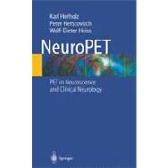
NeuroPET

by ; ;
  • ISBN13:

    9783540006916

  • ISBN10:

    3540006915

  • Edition: CD
  • Format: Hardcover
  • Copyright: 2004-08-01
  • Publisher: Springer Nature
  • Purchase Benefits
  • Free Shipping Icon Free Shipping On Orders Over $35!
    Your order must be $35 or more to qualify for free economy shipping. Bulk sales, PO's, Marketplace items, eBooks and apparel do not qualify for this offer.
  • eCampus.com Logo Get Rewarded for Ordering Your Textbooks! Enroll Now
List Price: $109.00 Save up to $89.20
  • Digital
    $42.90
    Add to Cart

    DURATION
    PRICE

Summary

Neurological applications of PET include using it to identify Alzheimer's Disease, including differentiating between other forms of Dementia, to show causes of seizures that were otherwise unclear, and to diagnose psychiatric disorders like Schizophrenia. Although other books on PET may have a chapter or section on neurological applications, this is the only comprehensive and up-to-date book on neurological PET. It reviews PET in neuroscience with particular emphasis on findings that indicate its potential for improving diagnosis and treatment in neurology and psychiatry. Improving the transfer of the huge scientific developments in brain PET into clinical care will produce tangible human benefit. To this end, NeuroPET focuses on practical and potentially clinically relevant issues and identify solid ground as well as open questions. In addition, the book includes major chapters on the methodological background, including tracer physiology and kinetic modeling, and a comprehensive literature review. Included in this extremely useful package is a CD-ROM that features video clips and interactive navigation in 3D data sets that illustrate the information contained in PET images in major neurological diseases and its potential to improve diagnosis and neurosurgical planning in brain tumors.

Table of Contents

1 Introduction 1(6)
2 Clinical Studies 7(136)
2.1 Dementia and Memory Disorders
7(25)
2.1.1 Clinical and Research Issues
7(2)
2.1.2 Alzheimer's Disease and Mild Cognitive Impairment
9(15)
2.1.2.1 Cerebral Glucose Metabolism
9(11)
2.1.2.2 Cholinergic Degeneration
20(2)
2.1.2.3 Other Receptors and Transmitters
22(1)
2.1.2.4 Amyloid Imaging
22(1)
2.1.2.5 Activation Studies
23(1)
2.1.3 Dementia with Lewy Bodies
24(1)
2.1.4 Frontotemporal Dementia and Related Disorders
25(3)
2.1.5 Vascular Dementia
28(2)
2.1.6 Creutzfeldt-Jakob Disease
30(1)
2.1.7 Other Memory Disorders
31(1)
2.1.7.1 Hypoxic and Global Ischemic Brain Damage
31(1)
2.1.7.2 Other Amnesic Syndromes
31(1)
2.2 Movement Disorders
32(31)
2.2.1 Idiopathic Parkinson Disease
35(13)
2.2.1.1 Diagnostic Issues
35(2)
2.2.1.2 Dopamine Synthesis
37(2)
2.2.1.3 Dopamine Release and Turnover
39(1)
2.2.1.4 Dopamine Transporters
39(1)
2.2.1.5 Vesicular Monoamine Transporter
40(1)
2.2.1.6 Dopamine Receptors
40(1)
2.2.1.7 Mesocortical Dopaminergic Innervation
41(1)
2.2.1.8 Other Transmitters
41(1)
2.2.1.9 Resting Glucose Metabolism
41(1)
2.2.1.10 Activation Studies
42(1)
2.2.1.11 Familial PD
42(2)
2.2.1.12 Disease Progression
44(1)
2.2.1.13 Depression in PD
45(1)
2.2.1.14 Improving and Understanding Treatment Effects
45(3)
2.2.2 Multiple System Atrophy
48(4)
2.2.2.1 Olivo-ponto-cerebellar Atrophy
49(1)
2.2.2.2 Striatonigral Degeneration
50(1)
2.2.2.3 Shy-Drager Syndrome
50(2)
2.2.3 Other Neurodegenerative Disorders
52(4)
2.2.3.1 Progressive Supranuclear Palsy
52(1)
2.2.3.2 Corticobasal Degeneration
53(2)
2.2.3.3 Disorders with Abnormal Deposition of Metals
55(1)
2.2.3.4 Parkinsonism Attributable to Toxic and Inflammatory Brain Damage
55(1)
2.2.3.5 Amyotrophic Lateral Sclerosis
56(1)
2.2.3.6 Other Rare Disorders
56(1)
2.2.4 Hyperkinetic Syndromes
56(4)
2.2.4.1 Huntington's Chorea (Huntington Disease)
56(2)
2.2.4.2 Other Choreic Disorders
58(1)
2.2.4.3 Restless Legs Syndrome
58(1)
2.2.4.4 Essential and Orthostatic Tremor
59(1)
2.2.4.5 Tourette Syndrome
59(1)
2.2.5 Dystonia and Related Disorders
60(1)
2.2.5.1 Idiopathic Dystonia and Torticollis
60(1)
2.2.5.2 DOPA-responsive Dystonia
61(1)
2.2.5.3 Dystonia Attributable to Localized Lesions or Degeneration
61(1)
2.2.6 Ataxia
61(3)
2.2.6.1 Spinocerebellar Ataxia
62(1)
2.2.6.2 Friedreich's Ataxia
63(1)
2.2.6.3 Other Ataxias
63(1)
2.3 Brain Tumors
63(1)
2.3.1 Biological Grading
64(22)
2.3.1.1 Glucose Consumption
64(4)
2.3.1.2 Amino Acid Uptake
68(3)
2.3.1.3 Nucleoside Uptake
71(1)
2.3.2 PET-Guided Stereotactic Biopsy
71(1)
2.3.3 Differentiation Between Recurrent Tumor and Radiation Necrosis
71(2)
2.3.4 Monitoring of Therapy
73(4)
2.3.5 Identification of Hypoxic Tumor Tissue
77(1)
2.3.6 Improving and Planning Therapy
77(2)
2.3.7 New Therapies
79(1)
2.3.8 Other Tracers for Brain Tumors
80(1)
2.3.9 Extracranial Tumors of the Nervous System
80(1)
2.3.10 Detection of Primary Tumors
81(1)
2.3.11 Activation Studies
81(5)
2.3.11.1 Motor Function
82(2)
2.3.11.2 Language
84(2)
2.4 Cerebrovascular Disease
86(14)
2.4.1 Ischemic Stroke
87(2)
2.4.2 Intracerebral and Subdural Hemorrhage
89(1)
2.4.3 Subarachnoid Hemorrhage
90(1)
2.4.4 Remote Effects (Diaschisis)
90(3)
2.4.5 Chronic Hemodynamic Impairment
93(2)
2.4.6 Moyamoya Disease
95(1)
2.4.7 Genetic Disorders (MELAS, CADASIL)
95(1)
2.4.8 Brain Function and Recovery After Stroke
95(5)
2.4.8.1 Aphasia
96(2)
2.4.8.2 Motor Function
98(1)
2.4.8.3 Neglect
99(1)
2.5 Epilepsy
100(13)
2.5.1 Localization of Epileptogenic Foci
101(8)
2.5.1.1 Temporal Lobe Epilepsy
101(4)
2.5.1.2 Neocortical Focal Epilepsy
105(1)
2.5.1.3 Ictal Studies
105(1)
2.5.1.4 GABA-A and Benzodiazepine Receptors
106(1)
2.5.1.5 Other Transmitters and Receptors
107(1)
2.5.1.6 Inflammatory Lesions and Glial Reaction
108(1)
2.5.2 Progression of Epileptic Lesions
109(1)
2.5.3 Prediction of Surgical Outcome
109(1)
2.5.4 Malformations of Cortical Development
109(2)
2.5.5 Childhood Epileptic Syndromes
111(1)
2.5.6 Language Dominance
112(1)
2.5.7 Effects of Surgical Intervention and Medical Treatment
112(1)
2.5.8 Generalized Epilepsy
113(1)
2.6 Other Neurological Disorders
113(10)
2.6.1 Traumatic Brain Injury
113(1)
2.6.2 Persistent Vegetative State
114(1)
2.6.3 Perinatal Brain Damage
115(2)
2.6.4 Inflammatory Disease
117(2)
2.6.5 Pain
119(2)
2.6.6 Migraine and Cluster Headache
121(1)
2.6.7 Narcolepsy
122(1)
2.6.8 Hypoparathyroidism (Fahr's Disease)
122(1)
2.6.9 Systemic Inherited Metabolic Disorders
122(1)
2.7 Psychiatric Disorders
123(29)
2.7.1 Depression
124(4)
2.7.1.1 Cerebral Blood Flow and Cerebral Metabolic Rate (Glucose)
125(2)
2.7.1.2 Serotonin Receptors
127(1)
2.7.1.3 Serotonin Transporters
128(1)
2.7.1.4 Other Transmitter Systems
128(1)
2.7.2 Fatigue
128(1)
2.7.3 Schizophrenia
129(2)
2.7.3.1 CBF and Energy Metabolism
129(1)
2.7.3.2 Activation Studies
129(1)
2.7.3.3 Dopamine Receptors and Dopamine Release
130(1)
2.7.3.4 Other Receptors
131(1)
2.7.3.5 Receptor Binding of Antipsychotic Drugs
131(1)
2.7.4 Anxiety Disorders
131(4)
2.7.4.1 Panic Disorder
132(1)
2.7.4.2 Phobic Disorders
132(1)
2.7.4.3 Obsessive-compulsive Disorder
133(1)
2.7.4.4 Posttraumatic Stress Disorder
134(1)
2.7.5 Drug and Substance Abuse
135(6)
2.7.5.1 Alcoholism
135(1)
2.7.5.2 Cocaine
136(2)
2.7.5.3 Amphetamine and Derivatives
138(1)
2.7.5.4 Opiates
139(1)
2.7.5.5 Nicotine
140(1)
2.7.5.6 Hallucinogens
140(1)
2.7.5.7 Phencyclidine
140(1)
2.7.6 Personality and Behavioral Disorders
141(1)
2.7.6.1 Borderline Personality Disorder
141(1)
2.7.6.2 Violence and Suicide
141(1)
2.7.7 Anorexia Nervosa and Bulimia
142(1)
3 Imaging Brain Function 143(1)
3.1 Blood-Brain Barrier Transfer
143(4)
3.2 Cerebral Blood Flow
147(2)
3.3 Oxygen Consumption
149(1)
3.4 Glucose Consumption
150(2)
3.5 Influence of Brain Function on CBF and Metabolism
152(35)
3.5.1 Wakefulness and Sleep
153(1)
3.5.2 Effect of Drugs
153(1)
3.5.3 The "Resting State"
154(1)
3.5.4 Activation Studies
155(1)
3.6 Tissue Oxygen Pressure and pH
156(1)
3.7 Amino Acid Transport and Protein Synthesis
156(1)
3.7.1 Transport-only Tracers
157(1)
3.7.2 Tracers with Incorporation into Proteins
158(2)
3.7.3 Precursors and Analogues for Transmitter Systems
160(1)
3.8 Nucleosides and DNA Synthesis
160(1)
3.9 Molecular Imaging
161(2)
3.9.1 Herpes Simplex Virus Thymidine Kinase Imaging
163(1)
3.9.2 Reporter Gene Imaging
164(1)
3.9.3 Oligonucleotides
164(1)
3.9.4 Apoptosis and p53
164(1)
3.9.5 Cell Trafficking
164(1)
3.9.6 Angiogenesis
165(1)
3.10 Dopamine System
165(8)
3.10.1 Precursors and Analogues of Dopamine
165(2)
3.10.2 Dopamine Transporter
167(1)
3.10.3 Vesicular Monoamine Transporter
168(1)
3.10.4 D1 Receptors
169(1)
3.10.5 D2 Receptors
169(1)
3.10.6 Studies of Synaptic Dopamine Release
170(2)
3.10.7 Monoamine Oxidase
172(1)
3.11 Cholinergic System
173(3)
3.11.1 Acetylcholine Synthesis
173(1)
3.11.2 Vesicular Acetylcholine Transporter
173(1)
3.11.3 Nicotinic Receptors
173(1)
3.11.4 Muscarinic Receptors
174(1)
3.11.5 Acetylcholine Esterase
174(2)
3.12 Serotonin System
176(4)
3.12.1 Serotonin Precursors
176(1)
3.12.2 Serotonin Transporter
177(1)
3.12.3 Serotonin Receptors
177(3)
3.12.3.1 Serotonin 1A Receptors
177(3)
3.12.3.2 Serotonin 2A Receptors
180(1)
3.13 Gamma-aminobutyric acid (GABA)
180(2)
3.13.1 Central Benzodiazepine-binding Sites
181(1)
3.13.2 Peripheral Benzodiazepine Receptors
181(1)
3.14 Glutamate and NMDA Receptors
182(1)
3.15 Adenosine Receptors
182(1)
3.16 Histamine Receptors
182(1)
3.17 Cannabinoid Receptors
183(1)
3.18 Opioid Receptors and Sigma Receptor
183(1)
3.19 Steroid Receptors
184(1)
3.20 Substance P
184(1)
3.21 Secondary Neurotransmitters
185(2)
4 Data Acquisition, Reconstruction, Modeling, Statistics 187(1)
4.1 Positron Emitters and Tracers
187(2)
4.2 Scanners and Detector Systems
189(2)
4.3 Data Acquisition
191(1)
4.4 Image Reconstruction
192(2)
4.5 Motion Detection and Correction
194(1)
4.6 Data Visualization
194(1)
4.7 Image Coregistration
195(40)
4.7.1 Fiducial Markers
196(1)
4.7.2 Matching of Brain Structures
197(1)
4.7.3 Algorithms for Maximization of Similarity
198(1)
4.7.4 PET/CT and PET/MRI Scanners
199(1)
4.8 Anatomical Standardization
200(1)
4.8.1 Orientation of Transaxial Slices
200(1)
4.8.2 Matching of Individual Brains to Image Templates ("Spatial Normalization")
201(1)
4.8.3 Atlases for Identification of Anatomical Structures
202(1)
4.9 Physiological Modeling
203(2)
4.9.1 Blood and Homogeneous Tissue (One Tissue Compartment)
205(7)
4.9.1.1 Transport across the BBB
206(1)
4.9.1.2 Cerebral Blood Flow
207(4)
4.9.1.3 Mixed Effects of Transport and Blood Flow
211(1)
4.9.1.4 Measurement of the Partition Coefficient
211(1)
4.9.1.5 Saturable Transport and Enzyme Reactions
212(1)
4.9.2 Metabolism
212(6)
4.9.2.1 Irreversible Metabolism (Metabolic Trapping)
213(1)
4.9.2.2 Measurement of Local Cerebral Glucose Metabolism
214(2)
4.9.2.3 The Reference Tissue Model for Dopamine Synthesis and Storage
216(1)
4.9.2.4 Reversible Metabolism or Binding
217(1)
4.9.3 Receptor Binding
218(4)
4.9.3.1 Irreversible Receptor Binding
219(1)
4.9.3.2 Equilibrium Approaches for Quantification of Reversible Receptor Binding
220(2)
4.9.4 Extraction of Model Parameters from Kinetic Data
222(1)
4.10 Quantitative Data Analysis
223(12)
4.10.1 Regions/Volumes of Interest
225(1)
4.10.2 Partial Volume Correction
226(1)
4.10.3 Statistical Models for Quantitative Data
227(4)
4.10.4 Parametric Imaging
231(1)
4.10.5 Statistical Parametric Mapping
232(1)
4.10.6 Lesion Detection and Pattern Recognition
233(2)
References 235(50)
Subject Index 285

Supplemental Materials

What is included with this book?

The New copy of this book will include any supplemental materials advertised. Please check the title of the book to determine if it should include any access cards, study guides, lab manuals, CDs, etc.

The Used, Rental and eBook copies of this book are not guaranteed to include any supplemental materials. Typically, only the book itself is included. This is true even if the title states it includes any access cards, study guides, lab manuals, CDs, etc.

Rewards Program