did-you-know? rent-now

Amazon no longer offers textbook rentals. We do!

did-you-know? rent-now

Amazon no longer offers textbook rentals. We do!

We're the #1 textbook rental company. Let us show you why.

9780683301229

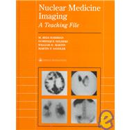
Nuclear Medicine Imaging A Teaching File

by ; ; ;
  • ISBN13:

    9780683301229

  • ISBN10:

    0683301225

  • Edition: 2nd
  • Format: Hardcover
  • Copyright: 1999-06-04
  • Publisher: LWW
  • Purchase Benefits
  • Free Shipping Icon Free Shipping On Orders Over $35!
    Your order must be $35 or more to qualify for free economy shipping. Bulk sales, PO's, Marketplace items, eBooks and apparel do not qualify for this offer.
  • eCampus.com Logo Get Rewarded for Ordering Your Textbooks! Enroll Now
List Price: $159.00

Summary

Nuclear Medicine Imaging: A Teaching File is a collection of approximately 450-500 cases grouped by organ system that residents can study to test themselves. Each case features images, a history, brief discussion, differential diagnosis and the diagnosis, allowing readers to evaluate an image as though they were in the reading room.

Table of Contents

Contributors xvii
Publisher's Foreword xix
Foreword xxi
Preface xxiii
Acknowledgments xxv
Endocrine Imaging
1(50)
Martin P. Sandler
William H. Martin
Edward B. Moody
Benign Follicular Adenoma
4(2)
Lingual Thyroid
6(1)
Follicular Carcinoma of the Thyroid with Widespread Metastases
7(3)
Thyroid Dyshormonogenesis
10(2)
Viral Thyroiditis
12(2)
Graves' Disease
14(3)
Autonomously Functioning Thyroid Nodule
17(4)
Thyroid Goiter with Mediastinal Extension
21(3)
Multinodular Goiter
24(1)
Ectopic Parathyroid Adenoma
25(3)
MEN IIB Syndrome
28(2)
Parathyroid Adenoma and Metastatic Gastrinoma in MEN I Syndrome
30(3)
Insulinoma of the Tail of the Pancreas
33(2)
Congenital Absence of Right Lobe of Thyroid with Compensatory Hypertrophy of Left Lobe
35(2)
Metastatic Pheochromocytoma in a Patient with MEN II
37(3)
Papillary Carcinoma of the Thyroid with Soft Tissue and Skeletal Metastases
40(4)
Parathyroid Adenoma
44(3)
Primary Aldosteronoma
47(2)
Diffuse Toxic Goiter (Graves' Disease) with a Coexistent Solitary Hypofunctioning Nodule
49(2)
Pulmonary Scintigraphy
51(94)
M. Reza Hobibian
Acute Pulmonary Emboli with Rapid Resolution
56(2)
Resolution of Acute Pulmonary Emboli with Minimal Residual
58(2)
Acute Pulmonary Emboli with Partial Resolution
60(1)
High Probability for PE in a Patient with Previous Pulmonary Emboli
61(2)
Pulmonary Arterial Hypertension, Recurrent Pulmonary Emboli
63(2)
Recurrent Pulmonary Emboli
65(3)
Massive Pulmonary Emboli Fleischner's Sign, Westmark's Sign
68(2)
High Probability for PE in a Patient with Parenchymal Infiltrate
70(2)
High Probability for Upper Lobe PE in a Patient with Parenchymal Infiltrate
72(3)
High Probability for PE in a Patient with COPD
75(2)
High Probability for PE in a Patient with Chest Mass
77(2)
Low Probability for PE in a Patient with Parenchymal Infiltrate
79(2)
Low Probability for PE in a Patient with Pleural Effusion
81(2)
Normal Perfusion Scan with Iatrogenic Microthrombus
83(1)
Low Probability for PE
84(3)
Stripe Sign
87(2)
Positive Radionuclide Lower Extremity Venogram
89(2)
Positive Red Blood Cell Labeled Venogram and PE
91(3)
Intermediate Probability for PE
94(2)
Triple Matched Defect Lower Lobe
96(3)
Intermediate Probability for PE
99(2)
Mucus Plug
101(2)
Respiratory Distress Syndrome
103(2)
Pulmonary Edema
105(2)
Hydropneumothorax
107(2)
Radiation Pneumonitis
109(2)
Fatty Infiltration of the Liver
111(2)
COPD
113(2)
COPD Reverse Mismatch
115(2)
Lower Lobe Emphysema, Alpha I Antitrypsin Deficiency
117(2)
Upper Lobe Bullous Disease
119(2)
Hilar Mass
121(2)
False-Positive High Probability for PE
123(3)
Tumor Microembolism
126(2)
Pulmonary Tumor Emboli
128(3)
Pulmonary Metastases
131(2)
Osler-Weber-Rendu Syndrome
133(4)
Bronchogenic Carcinoma---Surgical Candidate
137(2)
Gallium-67 Scan, Active Pulmonary Sarcoidosis
139(2)
Gallium-67 Scan, Pneumocystis Carinii Pneumonia
141(4)
Cardiovascular Imaging
145(72)
William H. Martin
Mark A. Coppess
Joao V. Vitola
Martin P. Sandler
Normal Stress 201Thallium Perfusion: Prognostic Implication
155(2)
Myocardial Ischemia due to Two-Vessel Disease
157(1)
Multivessel Ischemia: Pre-Operative Evaluation
158(1)
Lateral Wall Ischemia due to Left Circumflex Stenosis
159(1)
Myocardial Infarction: Pre-Op Assessment for Noncardiac Surgery
160(1)
Myocardial Ischemia; Utility of 24-Hour 201Thallium Imaging
161(2)
The Reverse Redistribution Defect
163(2)
Myocardial Viability Assessment Using FDG Spect
165(1)
Normal MIBI Perfusion: Advantages of MIBI Spect
166(1)
Ischemia due to LAD Stenosis
167(1)
Rest FDG/MIBI SPECT: Viability Assessment
168(2)
Ischemic Cardiomyopathy: FDG/MIBI SPECT
170(1)
Extensive Myocardial Scarring: Gated SPECT
171(2)
False Positive Perfusion Spect due to LBBB
173(2)
Soft Tissue Attenuation Artifacts
175(3)
Radionuclide Ventriculography: Dilated Cardiomyopathy
178(2)
Quantitation of Ischemia Using Circumferential Profiling
180(2)
Gastrointestinal Artifacts
182(2)
Quantitation of Ischemia Using the CeQUEL Program
184(2)
Positron Emission Tomography (PET) for Post-PTCA Ischemia
186(1)
Extracardiac (Puerperal Breast) Uptake of MIBI
187(1)
RVG: Extracardiac Uptake of 99mTc-RBC
188(2)
RVG: Technical Problems
190(1)
Infarct-Avid Imaging
191(4)
Doxorubicin-Induced Left Ventricular Dysfunction
195(1)
Pulmonary Uptake of 201Thallium
196(1)
Lateral Ischemia: Advantages of 201Thallium PECT
197(1)
Planar Perfusion Imaging
198(1)
RVG: Diastolic Dysfunction
199(2)
Ischemia Demonstrated by Exercise RVG
201(2)
Acute Ischemic Syndromes: Rest Perfusion SPECT
203(3)
Post-PTCA Perfusion SPECT
206(3)
Primary Pulmonary Hypertension
209(1)
LBBB: Stress Perfusion Imaging
210(3)
Multivessel CAD: Stress FDG/MIBI SPECT
213(4)
Neurologic Imaging
217(56)
Dominique Delbeke
Brain Flow Present (99mTc-Gllucoheptonate)
223(1)
Interictal Seizure Focus in the Right Temporal Lobe (99mTc-HMPAO, 18F-FDG)
224(2)
Advanced Alzheimer's Disease (18F-FDG)
226(2)
Brain Flow Absent (99mTc-Gllucoheptonate, Technical Considerations)
228(2)
CNS Lymphoma in an HIV Patient (18F-FDG)
230(1)
Low-Grade Tumor (18F-FDG)
231(1)
High-Grade Tumor (18F-FDG)
232(1)
Pick's Disease (18F-FDG)
233(1)
Recurrent High-Grade Tumor (18F-FDG)
234(1)
Craniopharyngioma (18F-FDG)
235(2)
Ischemia in the Distribution of the Left Carotid Artery and Infarct of the Right Occipital Cortex (99mTc-HMPAO with Acetazolamide Challenge)
237(2)
Normal Cisternogram with Pledgets Positive for a CSF Leak (111In-DTPA)
239(3)
Normal Cerebral Perfusion During Balloon Occlusion Test of the Right Carotid Artery (99mTc-HMPAO)
242(1)
Low CSF Pressure Headache (11In-DTPA)
243(3)
Huntington's Disease (18F-FDG)
246(1)
Decreased Perfusion to the Right Hemisphere During Balloon Occlusion Test of the Right Carotid Artery (99mTc-HMPAO)
247(1)
Atypical Alzheimer's Disease (18F-FDG)
248(1)
High-Grade Tumor with Adjacent Ictal Seizure Focus (18F-FDG)
249(2)
Infarct of the Left Anterior Cerebral Artery as a Complication of Surgical Clipping of an Aneurysm of the Anterior Communicating Artery (99mTc-HMPAO)
251(2)
Ictal Seizure Focus in the Right Mesial Frontal Lobe (99mTC-HMPAO, 18F-FDG)
253(3)
Aspergilloma (18F-FDG)
256(1)
Ictal Seizure Focus Arising from a Region of Cortical Dysplasia with Possible Closed Lip Schizencephalic Cleft (99mTc-HMPAO, 18F-FDG)
257(3)
Multi-Infarct Dementia (18F-FDG)
260(2)
Normal Pressure Hydrocephalus (111In-DTPA)
262(2)
Herpes Encephalitis (99mTc-glucoheptonate, (18F-FDG)
264(2)
High-Grade Tumor Recurrence (201T1)
266(2)
Occluded Ventriculoperitoneal Shunt (99mTc-DTPA)
268(2)
Radiation Necrosis (18F-FDG)
270(3)
Gastrointestinal Scintigraphy
273(92)
M. Reza Hobibian
Cirrhosis on Liver Scan
278(2)
Hepatoma Superimposed on Cirrhosis
280(2)
Disseminated Liver Metastases
282(2)
Liver Abscess
284(1)
End-stage Hepatic Disease
285(3)
Atlas Review
288(4)
Hepatocellular Carcinoma
292(1)
Focal Fatty Infiltration vs. Tumors (Fat)
293(2)
Focal Fatty Infiltration vs. Tumor (Tumor)
295(2)
Liver Mass
297(3)
Cavernous Hemangioma
300(3)
Cavernous Hemangioma with SPECT
303(3)
False Positive Scan due to Ascites
306(2)
Renal Uptake of 99mTc-Sulfur Colloid
308(3)
Accessory Spleen
311(2)
Space Occupying Lesion of the Spleen
313(3)
Malignant Liver Mass, Multi-Image Studies
316(3)
Focal Nodular Hyperplasia
319(3)
Liver Carcinoid
322(2)
Recurrent Colon Carcinoma
324(3)
Segmental Biliary Obstruction
327(2)
GI Bleeding---Small Bowel
329(2)
GI Bleeding---Large Bowel
331(2)
Abdominal Blood Pool Image
333(4)
Hepatic Fibrosis
337(1)
Renal Tumor
338(1)
Left Renal Artery Pseudoaneurysm
339(2)
Rim Sign
341(2)
Morphine Augmentation
343(2)
Gastric Reflux
345(2)
Cystic Duct Remnant
347(1)
Biliary Leak
348(2)
Cholesytojejunostomy Anastomosis
350(1)
Partial Biliary Obstruction
351(2)
Common Bile Duct Obstruction
353(2)
Chronic Cholecystitis
355(2)
Pseudomembraneous Colitis
357(1)
Right Lower Quadrant Abscess
358(1)
Hepatic Hydrothorax
359(1)
LaVeen Shunt
360(2)
Warthin's Tumor
362(3)
Renal Scintigraphy
365(70)
Sadrollah Bazarjani
Dominique Delbeke
Normal Scrotal Scintigram
370(2)
Early Testicular Torsion
372(3)
Late Testicular Torsion with Acute Hemorrhagic Necrosis
375(2)
Epididymo-Orchitis
377(2)
Unilateral Vesicoureteral Reflux
379(2)
Bilateral Vesicoureteral Reflux
381(1)
Indirect Radionuclide Cystogram
382(1)
Pyelonephritis
383(3)
Unilateral Renovascular Hypertension
386(4)
Bilateral Renovascular Hypertension
390(3)
Normal 99mTc DTPA Renal Scintigram
393(3)
Normal 99mTc MAG3 Renogram
396(2)
Acute Renal Failure (with 99mTc DTPA)
398(2)
Acute Renal Failure (with 99mTc MAG3)
400(2)
Non-Obstructive Hydronephrosis
402(2)
Obstructive Hydronephrosis
404(3)
Ectopic Pelvic Kidney on Bone Scan
407(2)
Normal Transplant Renogram
409(2)
Acute Renal Transplant Rejection
411(2)
Vascular Complication of the Transplanted Kidney
413(2)
Posttransplant Lymphocele
415(2)
Urinary Extravasation
417(2)
Non-Functioning Kidney
419(2)
Gallbladder Visualization with 99mTc MAG3
421(1)
Residual Function in Old Renal Transplant
422(2)
Malignant Tumor within a Renal Allograft
424(2)
Prominent Column of Bertin
426(1)
Duplex System
427(3)
Unilateral Hydronephrosis
430(1)
Renal Cell Carcinoma
431(4)
Skeletal Scintigraphy
435(184)
M. Reza Hobibian
Solitary Sclerotic Metastases
445(2)
Solitary Lytic Metastases
447(2)
Hot and Cold Metastases
449(2)
Photopenic Metastases
451(1)
Renal Mass with Metastases
452(2)
The Solitary Rib Lesion
454(3)
Progressive Metastases Versus Flare
457(3)
Superscan
460(3)
Appendicular Metastasis---Ulna
463(2)
Appendicular Metastasis---Tibia
465(2)
Metastasis of a Doughnut Appearance
467(3)
Metastases and Paget's Disease
470(1)
Resolving Metastases, Postchemotherapy
471(2)
Metastases to the Bone and Liver
473(4)
Metastases to the Bone, Liver, and Kidney
477(2)
Metastases from Thyroid Carcinoma
479(3)
Osteogenic Sarcoma Metastasized to the Lungs
482(2)
Bone Destruction and Soft Tissue Mass
484(3)
Metastases Mimicking Paget's Disease
487(1)
Carcinoid Metastases
488(2)
Chondrosarcoma
490(2)
Osteochondroma
492(2)
Osteoid Osteoma
494(4)
Fibrous Dysplasia
498(6)
Non-Osteogenic Fibroma
504(3)
Enchondroma
507(1)
Benign Chondroblastoma
508(1)
Ribbing's Disease
509(2)
Bone Island
511(2)
False-Negative Scan
513(2)
Polyostotic Paget's Disease
515(3)
Subradiographic Paget's Disease
518(2)
Sacral Paget's
520(2)
Renal Osteodystrophy
522(3)
End-Stage Renal Disease
525(2)
Renal Osteodystrophy-Amyloidoma
527(2)
Osteomyelitis due to Penetrating Wound
529(3)
Osteomyelitis of Metatarsal Bone
532(2)
Osteomyelitis of Greater Trochanter
534(2)
Diskitis
536(2)
Cold Defects on 111In Leukocyte Images
538(3)
Charcot Arthropathy
541(2)
Rhabdomyolysis
543(2)
Myositis Ossificans
545(2)
Polymyositis
547(2)
Longitudinal Stress Fracture
549(2)
Pathological Fracture
551(2)
Stress Fracture
553(2)
Stress Fracture and Shin Splint
555(3)
Stress Fracture of Cancellous Bone
558(4)
Occult Fracture of Femur
562(2)
Occult Fracture of Sacrum
564(2)
Fracture of the Scaphoid, Avascular Necrosis Lunate
566(2)
Fracture of Sesamoid and Navicular Bone
568(3)
Fracture of Patella
571(3)
Non-Union Fracture
574(3)
Reflex Sympathetic Dystrophy
577(2)
Idiopathic Regional Migratory Osteopenia
579(2)
Reflex Sympathetic Dystrophy
581(2)
Avascular Necrosis of Femoral Head
583(2)
Osteochondritis Dissecans
585(2)
Freiberg's Disease
587(1)
Vascular Pseudotumor
588(2)
Uterine Hyperemia
590(2)
Reno-Colonic Fistula
592(2)
Temporomandibular Joint Arthritis
594(2)
Mandibular Bone Graft
596(1)
Hypertrophic Osteoarthropathy
597(2)
Sickle Cell Disease
599(3)
Myocardial Uptake
602(2)
Ankylosing Spondylitis
604(3)
Osteoporotic Vertebral Fracture
607(2)
Postcardiac Resuscitation
609(1)
Gauntlet's Image
610(1)
Atlas Review
611(8)
Oncologic Imaging
619(104)
William H. Martin
Dominique Delbeke
M. Reza Habibian
Hodgkin's Lymphoma
625(2)
Recurrent Hodgkin's Disease
627(4)
Physiologic Thymic Rebound
631(4)
Recurrent Hodgkin's Disease, Post-Bone Marrow Transplantation
635(2)
Widespread Lymphoma
637(3)
Metastatic Colorectal Carcinoma Demonstrated by FDG-PET Imaging
640(3)
Colorectal Carcinoma Metastatic to Lung and Periaortic Nodes
643(2)
Colon Carcinoma with Hepatic and Nodal Metastases
645(3)
Focal Nodular Hyperplasia and Lactating Breasts
648(3)
Metastatic Carcinoid Tumor
651(2)
Cat Scratch Disease of the Liver
653(2)
Hepatic Arterial Perfusion Scintigraphy
655(4)
Monitoring Chemoembolization of a Hepatocellular Carcinoma with FDG-PET
659(2)
Metastatic Hepatocelluar Carcinoma
661(5)
Adrenal Metastasis
666(2)
Adrenocortical Carcinoma
668(2)
Cystic Adenocarcinoma of the Pancreas and Paget's Disease of the Scapula
670(3)
Recurrent Pancreatic Carcinoma with Widespread Metastases
673(4)
Pelvic Inflammatory Pseudotumor
677(3)
Metastases from Ovarian Carcinoma Identified by FDG-PET
680(2)
Malignant Solitary Pulmonary Nodule
682(3)
Bronchogenic Carcinoma with Mediastinal Lymphadenopathy
685(1)
Malignant Left Lung Nodule and Renal Transplant
686(1)
Recurrent Bronchogenic Carcinoma with Artifacts
687(2)
Melanoma: Lymphoscintigraphy and Sentinel Node Localization
689(2)
Breast Cancer with Localization of Axillary Sentinel Node
691(2)
Widespread Melanoma Unsuspected Clinically
693(1)
Breast Carcinoma with Axillary Metastasis
694(2)
Metastatic Breast Carcinoma
696(2)
Small Cell Lung Cancer with Extensive Metastases
698(2)
Metastatic Medullary Carcinoma of the Thyroid
700(2)
Gastrinoma, Somatostatin-Receptor Positive
702(4)
Ectopic ACTH Syndrome due to Metastatic Bronchial Carcinoid
706(3)
Metastatic Neuroblastoma
709(3)
Carotid Paraganglioma
712(2)
Melanoma, Metastasis to the Bowel Wall and Adrenal Gland
714(3)
Soft-Tissue Sarcoma with Metastasis
717(6)
Infectious Process
723(62)
Edwin L. Born Jr.
William H. Martin
Osteomyelitis of Foot in a Diabetic Patient
726(3)
Charcot Arthropathy in a Diabetic Patient
729(4)
Miliary Tuberculosis
733(2)
Chronic Aorto-Bifemoral Bypass Graft Infection
735(3)
Acute Femoral-Femoral Bypass Graft Infection
738(2)
Altered Biodistribution of 67Ga Following Multiple Blood Transfusions
740(2)
Infected AV Dialysis Shunt
742(1)
FUO in Patient with Lymphoma
743(3)
Infected Peritoneal Dialysis Tunnel Catheter
746(2)
Discitis of Lumbar Spine
748(2)
Multiple Liver Abscesses
750(3)
Brain Abscess
753(2)
Decreased Leukocyte Uptake in Bone Marrow Following Radiation Therapy
755(1)
Aortic Graft Infection with 99mTc-HMPAO
756(3)
Pain in HIP Prosthesis: Infection and Loosening
759(2)
Bowel Infarct
761(1)
Pseudomembranous Colitis and Polycystic Liver
762(1)
Acute Pyelonephritis (Unilateral)
763(1)
PCP in an AIDS Patient
764(2)
Abdominal Abscess following Appendectomy
766(2)
Reiter's Syndrome
768(2)
Three Phase Bone Scans of Osteomyelitis and Cellulitis
770(2)
Sinusitis in Patient with Wegener's Granulomatosis
772(2)
Pulmonary Uptake of Radiolabeled Leukocytes
774(2)
Thoracic Vertebral Instability without Infection and Vertebral Osteomyelitis
776(3)
Inflammatory Bowel Disease
779(1)
Lemierre's Syndrome
780(5)
Pediatric Nuclear Imaging
785(66)
Susan Passalaqua
Dominique Delbeke
Langerhans Cell Histiocytosis
788(3)
Lingual Thyroid
791(2)
Horseshoe Kidney
793(1)
Autosomal Recessive Polycystic Kidney Disease
794(2)
Vesicoureteral Reflux
796(2)
Meckel's Diverticulum
798(2)
Neuroblastoma
800(2)
Biliary Atresia
802(2)
Multicystic Dysplastic Kidney
804(3)
Osteoid Osteoma
807(2)
Gastroesophageal Reflux
809(1)
Choledochal Cyst
810(1)
Ewing's Sarcoma
811(2)
Osteosarcoma
813(4)
Lymphoma
817(3)
Testicular Torsion
820(3)
Acute Pyelonephritis
823(1)
Sicke Cell Disease
824(2)
Legg-Calve-Perthes Disease
826(2)
Fibrous Dysplasia
828(2)
Ureteropelvic Junction Obstruction
830(2)
Spiral Fracture of the Tibia
832(1)
Rhabdomyosarcoma
833(1)
Skeletal Lymphoma
834(3)
Appendicitis
837(1)
Renal Ectopia
838(2)
Osteomyelitis
840(3)
Ligamentous Avulsion
843(3)
Spondylolysis
846(2)
Non-Accidental Trauma
848(3)
Artifacts
851(38)
James A. Patton
Interference from a High-Energy External Source
856(1)
Photomultiplier Tube Drift (Obvious)
857(1)
Photomultiplier Tube Drift (Subtle)
858(2)
Pulse Height Analyzer (PHA) Window Off Peak
860(3)
Clothing Artifact
863(1)
Prosthetic Device Artifact
864(1)
Improper PHA Window and Improper Collimator
865(3)
Septal Penetration of Collimator
868(3)
Improper Injection Technique
871(2)
Urine Contamination
873(1)
Misadministration
874(1)
Patient with Right-to-Left Shunt
875(1)
Aerosol Contamination
876(1)
Center-of-Rotation Error in SPECT
877(1)
Improper Injection Technique
878(1)
Radiation Therapy Effects
879(1)
Damaged Collimator
880(1)
Intermittent Failure of Orientation Circuitry
881(1)
Double Exposure
882(1)
Patient with Hyperparathyroidism
883(1)
Free 99mTc Pertechnetate in Gastric Reflux Study with 99m Tc Sulfur Colloid
884(1)
Clumping of White Blood Cells in Survey for Infection
885(1)
Misadminitration: 99mTc Pertechnetate Instead of 99mTc HDP
886(1)
Infiltration Appearing in Lymph Node of 99mTc HDP Scan
887(1)
Free 99mTc Pertechnetate in 99mTc-HMPAO Brain SPECT
888(1)
Subject Index 889

Supplemental Materials

What is included with this book?

The New copy of this book will include any supplemental materials advertised. Please check the title of the book to determine if it should include any access cards, study guides, lab manuals, CDs, etc.

The Used, Rental and eBook copies of this book are not guaranteed to include any supplemental materials. Typically, only the book itself is included. This is true even if the title states it includes any access cards, study guides, lab manuals, CDs, etc.

Rewards Program