did-you-know? rent-now

Amazon no longer offers textbook rentals. We do!

did-you-know? rent-now

Amazon no longer offers textbook rentals. We do!

We're the #1 textbook rental company. Let us show you why.

9780750305914

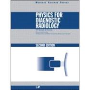
Physics for Diagnostic Radiology, Second Edition

by ;
  • ISBN13:

    9780750305914

  • ISBN10:

    0750305916

  • Edition: 2nd
  • Format: Nonspecific Binding
  • Copyright: 1999-05-01
  • Publisher: CRC Press
  • Purchase Benefits
  • Free Shipping Icon Free Shipping On Orders Over $35!
    Your order must be $35 or more to qualify for free economy shipping. Bulk sales, PO's, Marketplace items, eBooks and apparel do not qualify for this offer.
  • eCampus.com Logo Get Rewarded for Ordering Your Textbooks! Enroll Now
List Price: $74.95

Summary

This textbook is a complete course for radiologists studying for the FRCR part one exam, physicists and radiographers on specialised graduate courses in diagnostic radiology, or other appropriate undergraduate degrees. It follows the guidelines issued by the European Association of Radiology for training.It is a comprehensive, compact primer, appropriate to a teaching course some 50 hours in length. Its analytical approach deals in logical order with the wide range of imaging techniques available, and explains how to use imaging equipment.It includes the background physics necessary to understand the production of digitised images, nuclear medicine, and magnetic resonance imaging, It is assumed that students will be familiar with high school physics.It features extensive cross-referencing, practical analogies, further reading at the end of each chapter, exercises, exercises and a comprehensive selection of multiple choice questions (with answers) to test and reinforce student's understanding of the material presented.Major new features include: detailed discussion of patient dose; the use of digital radiography; image qual

Table of Contents

List of Contributors
vii
Acknowledgments viii
Introduction 1(2)
Fundamentals of Radiation Physics and Radioactivity
3(17)
Structure of the atom
3(1)
Nuclear stability and instability
3(3)
Radioactive concentration and specific activity
6(1)
Radioactive decay processes
6(1)
Exponential decay
7(1)
Half-life
8(1)
Secular and transient equilibrium
9(2)
Biological and effective half-life
11(1)
Gamma radiation
11(1)
X-rays and gamma rays as forms of electromagnetic radiation
12(1)
Quantum properties of radiation
13(1)
Inverse square law
13(1)
Interaction of radiation with matter
14(2)
Linear energy transfer
16(1)
Summary of energy changes in radiological physics
16(4)
Production of X-Rays
20(39)
Introduction
20(1)
The X-ray spectrum
20(6)
The continuous spectrum
20(2)
The low and high energy cut-off
22(1)
Shape of the continuous spectrum
23(1)
Line or characteristic spectra
23(1)
Factors affecting the X-ray spectrum
24(2)
Components of the X-ray tube
26(10)
The cathode
26(1)
The anode material
27(1)
Anode design
28(1)
Electrical circuits
29(4)
The tube envelope and housing
33(1)
Switching and timing mechanisms
34(1)
Electrical safety features
35(1)
Spatial distribution of X-rays
36(3)
Rating of an X-ray tube
39(10)
Introduction
39(1)
Electrical rating
40(2)
Thermal rating---considerations at short exposures
42(3)
Overcoming short-exposure rating limits
45(1)
Multiple or prolonged exposures
46(2)
Falling load generators
48(1)
Safety interlocks
49(1)
X-ray tube lifetime
49(1)
Mobile X-ray generators
49(3)
Single phase full wave rectified generators
50(1)
Constant potential generators
50(1)
Capacitor discharge units
51(1)
Quality assurance of performance for standard X-ray sets
52(3)
Summary
55(4)
Interaction of X-rays and Gamma Rays with Matter
59(24)
Introduction
59(1)
Experimental approach to beam attenuation
59(3)
Introduction to the interaction processes
62(1)
Bound and free electrons
62(1)
Attenuation, scatter and absorption
63(1)
The interaction processes
63(7)
Elastic scattering
63(1)
Photoelectric effect
63(2)
Compton effect
65(4)
Pair production
69(1)
Combining interaction effects and their relative importance
70(2)
Absorption edges
72(1)
Broad beam and narrow beam attenuation
73(2)
Filtration and beam hardening
75(4)
Conclusions
79(4)
The Image Receptor
83(31)
Introduction---band structure in solids
83(1)
Fluorescence, phosphorescence and thermoluminescence
84(2)
Phosphors and fluorescent screens
86(2)
Properties of phosphors
86(2)
Production of fluorescent screens
88(1)
Film--phosphor combinations in radiography
88(1)
X-ray film
88(5)
Film construction
88(1)
Characteristic curve and optical density
89(3)
Film gamma and film speed
92(1)
Latitude
93(1)
Film used with a fluorescent screen
94(1)
Cassettes
95(1)
Reciprocity
96(1)
Film--screen unsharpness
96(1)
Digital radiography
97(1)
Read out process
97(1)
Properties
97(1)
Eye--phosphor combination in fluoroscopy
98(1)
Phosphors used with image intensifiers
99(4)
Construction and mode of operation
99(3)
Quantum mottle
102(1)
Factors affecting the image
103(1)
The vidicon camera
103(2)
Cinefluorography
105(2)
Spot films
107(1)
Quality control of recording media and image intensification systems
107(3)
Summary
110(4)
The Radiological Image
114(21)
Introduction---the meaning of image quality
114(1)
The primary image
114(1)
Contrast
115(3)
Contrast on a fluorescent screen
116(1)
Contrast on a radiograph
116(1)
Origins of contrast for real and artificial media
117(1)
Effects of overlying and underlying tissue
118(1)
Reduction of contrast by scatter
119(1)
Variation in scatter with photon energy
119(1)
Reduction of scatter
120(2)
Careful choice of beam parameters
120(1)
Orientation of the patient
121(1)
Compression of the patient
121(1)
Use of grids
121(1)
Air gap technique
121(1)
Design of intensifying screen and film holder
121(1)
Grids
122(4)
Construction
122(1)
Use of grids
123(2)
Movement of grids
125(1)
Resolution and unsharpness
126(3)
Geometric unsharpness
126(1)
Patient unsharpness
127(1)
Combining unsharpnesses
128(1)
Geometric relationship of film, patient and X-ray source
129(2)
Magnification without distortion
129(1)
Distortion of shape and/or position
130(1)
Review of factors affecting the radiological image
131(4)
Choice of tube kilovoltage
131(1)
Exposure time
131(1)
Focal spot size
131(1)
Quality of anode surface
131(1)
Tube current
131(1)
Beam size
132(1)
Grids
132(1)
Focus--film and object--film distance
132(1)
Contrast enhancement
132(1)
Films and screens
132(1)
Film processing
132(3)
Radiation Measurement and Doses to Patients
135(28)
Introduction
135(1)
Ionization in air as the primary radiation standard
136(1)
The ionization chamber
137(2)
The Geiger--Muller counter
139(3)
The Geiger--Muller tube
140(1)
Comparison of ionization chambers and Geiger--Muller counters
141(1)
Relationship between exposure and absorbed dose
142(1)
Conversion of exposure in air to dose in air
142(1)
Conversion of dose in air to dose in tissue
143(1)
Practical radiation monitors
143(3)
Secondary ionization chambers
143(2)
Dose--area product meters
145(1)
Pocket exposure meters for personnel monitoring
146(1)
Thermoluminescent dosimeters (TLDs)
146(1)
Scintillation detectors and photomultiplier tubes
147(1)
Spectral distribution of radiation
148(1)
Semiconductor detectors
149(2)
Photographic film
151(1)
Variation of detector sensitivity with photon energy
152(1)
Patient doses in diagnostic radiology
153(6)
Principles
153(3)
Entrance doses in plain film radiography
156(1)
Entrance doses in fluoroscopy examinations
157(2)
Doses to organs
159(1)
Patient dose reduction in diagnostic radiology
159(1)
Technical factors
160(1)
Non-technical factors
160(1)
Conclusions
160(3)
Diagnostic Imaging with Radioactive Materials
163(29)
Introduction
163(1)
Principles of imaging
164(9)
The gamma camera
166(5)
Variations on the standard camera
171(2)
Factors affecting the quality of radionuclide images
173(9)
Information in the image and signal-to-noise ratio
173(1)
Choice of radionuclide
174(3)
Choice of radiopharmaceutical
177(1)
Performance of the imaging device
177(4)
Data display
181(1)
Dynamic investigations
182(5)
Data analysis
182(4)
Camera performance at high count rates
186(1)
Quality standards, quality assurance and quality control
187(2)
Radionuclide calibrators and accuracy of injected doses
187(1)
Gamma camera and computer
188(1)
Summary
189(3)
Assessment and Enhancement of Image Quality
192(27)
Introduction
192(1)
Factors affecting image quality
192(1)
Analogue and digital images
193(1)
Operation of the visual system
194(3)
Response to different light intensities
194(1)
Rod and cone vision
194(1)
Relationship of object size, contrast and perception
195(1)
Eye response to different spatial frequencies
196(1)
Limitations of a subjective definition of contrast
196(1)
Physical definition of contrast---signal-to-noise ratio
197(1)
Quantum noise
198(2)
Digital radiography (principles)
200(3)
Assessment of image quality
203(7)
Modulation transfer function
203(4)
Physical/physiological assessment
207(3)
Receiver operator characteristic (ROC) curves
210(5)
Principles
210(2)
ROC curves in practice
212(3)
Design of clinical trials
215(1)
Conclusions
216(3)
Special Radiographic Techniques
219(30)
Introduction
219(1)
High voltage radiography
219(3)
Macroradiography
222(2)
Digital radiography (practical detail)
224(6)
Introduction
224(1)
Methods of image formation
225(4)
Resolution requirements
229(1)
Patient doses
230(1)
Subtraction techniques
230(3)
Digital subtraction angiography
231(1)
Dual energy subtraction
232(1)
Time interval differencing
232(1)
Mammography
233(5)
Optimum kilovoltage and tube design
234(1)
Molybdenum anode tubes
234(1)
Tungsten anode tubes
235(1)
Film--screen combinations
235(2)
Patient doses
237(1)
Contrast improvement
237(1)
Quality control
237(1)
Xeroradiography
238(3)
Mode of use
239(1)
Formation of powder image
239(2)
Advantages and disadvantages of xeroradiography
241(1)
Paediatric radiology
241(2)
Dental radiology
243(6)
Intra-oral radiography
245(1)
Panoramic radiography
245(1)
Receptors
245(1)
Protection
246(1)
Quality assurance
246(3)
Tomographic Imaging
249(30)
Introduction
249(1)
Longitudinal tomography
250(4)
The linear tomograph
250(1)
Thickness of plane of cut
251(2)
Miscellaneous aspects of longitudinal tomography
253(1)
Computed axial transmission tomography
254(18)
Principles
254(1)
Data collection
255(3)
Data reconstruction
258(6)
Data presentation and storage
264(1)
System performance and quantum mottle
265(1)
Patient doses
266(3)
Spiral CT
269(1)
Artefacts
270(1)
Quality control
271(1)
Single photon emission computed tomography (SPECT)
272(2)
Positron emission tomography
274(2)
Conclusions
276(3)
Radiobiology and Radiation Risks
279(27)
Introduction
279(1)
Radiation sensitivity of biological materials
280(3)
Evidence for high radiosensitivity
280(1)
Cells particularly at risk; `law' of Bergonie and Tribondeau
280(1)
Time course of radiation-induced death
281(1)
Other mechanisms of radiation-induced death
281(2)
Evidence on radiobiological damage from cell survival curve work
283(4)
Cellular recovery and dose rate effects
283(3)
Radiobiological effectiveness (RBE)
286(1)
Radiation weighting factors, equivalent dose and the sievert
287(1)
Radiation effects on humans
288(5)
Deterministic and stochastic effects
288(1)
Carcinogenesis
289(1)
Mutagenesis
290(3)
Risk factors and collective doses
293(2)
Risks from radiological examinations
295(2)
Tissue weighting factors and effective dose
295(1)
Effective doses and risks in radiology
296(1)
Radiation risks in context
296(1)
Special high-risk situations---irradiation of children or in utero
297(3)
Lethal effects
297(1)
Malformations and other developmental effects
298(1)
Radiation effects on the developing brain
298(1)
Cancer induction
299(1)
Pre-conception risk (leukaemia clusters)
299(1)
Hereditary effects
299(1)
Summary---current thinking on risks of foetal irradiation (NRPB 1993b)
300(1)
Worked example on risk associated with an abdominal examination during undeclared pregnancy
300(2)
Basic data on good radiographic set-up
300(1)
Calculation
300(1)
Risk assessment
301(1)
Notes
301(1)
Risk from ingested or injected activity
302(1)
Summary
302(4)
Practical Radiation Protection and Legislation
306(24)
Introduction
306(1)
Role of radiation protection in diagnostic radiology
306(4)
Principles of protection
306(1)
Patient protection
307(2)
Staff protection
309(1)
Public protection
309(1)
European and UK legislation
310(8)
Introduction
310(1)
European legislation
310(1)
Basic Safety Standards Directive 96/29/Euratom (1996)
310(1)
Medical Exposures Directive 97/43/Euratom (1997)
311(1)
Outside Workers Directive 90/641/Euratom (1990)
312(1)
UK legislation
313(1)
Radioactive Substances Act 1993
313(1)
The Medicines (Administration of Radioactive Substances) Regulations 1978
313(1)
The Ionising Radiations (Protection of Persons Undergoing Medical Examination or Treatment) Regulations 1988
314(1)
Radioactive Material (Road Transport) Great Britain Regulations 1996
315(1)
Ionising Radiations Regulations 1985
315(3)
The Ionising Radiations Regulations (Outside Workers) 1993
318(1)
New UK legislation
318(1)
Doses to staff and patients
318(2)
Staff doses
318(1)
Patient doses
319(1)
X-ray rooms
320(1)
Introduction
320(1)
Points of note on room design
320(1)
Nuclear medicine
321(2)
Introduction
321(1)
Annual limit on intake
321(1)
Derived limits for airborne and surface contamination
322(1)
Special precautions in nuclear medicine
322(1)
Personal dosimetry
323(7)
Range of response
323(1)
Linearity of response
323(1)
Calibration against radiation standards
323(1)
Variation of sensitivity with radiation energy
323(1)
Sensitivity to temperature and humidity
323(1)
Uniformity of response within batches
324(1)
Maximum time of use
324(1)
Compactness
324(1)
Permanent visual record
324(1)
Indication of type of radiation
324(1)
Indication of pattern of radiation
325(1)
Cost
326(1)
Electronic personal dosimeters
326(4)
Diagnostic Ultrasound
330(48)
Introduction
330(1)
Ultrasound waves and their properties
331(5)
Longitudinal and transverse waves
331(1)
Excess pressure and frequency
332(1)
Speed of sound
332(1)
Wavelength
332(1)
Characteristic acoustic impedance
333(1)
Energy, power and intensity
333(1)
Decibels
334(1)
Pulse waves, energy spectra and bandwidth
335(1)
The propagation of ultrasound waves in tissue
336(3)
Speed of sound
336(1)
Attenuation
336(1)
Attenuation coefficient
337(1)
Reflection
337(2)
Refraction
339(1)
Scattering
339(1)
The ultrasound transducer
339(2)
Ultrasound beams
341(2)
Ultrasonic imaging modes
343(2)
Pulse--echo range finding
343(1)
A-mode scans
344(1)
M-mode scans
344(1)
B-mode scans
345(1)
Doppler imaging
345(1)
B-mode scanners
345(19)
Factors that are important for the quality of a B-mode image
345(3)
Beam forming
348(1)
Single element mechanical sector scanners
348(1)
Annular array mechanical sector scanners
348(3)
Linear and curvilinear array scanners
351(2)
Phased array scanners
353(1)
Intra-corporeal probes (endoprobes)
354(1)
Write zoom
355(1)
Amplifying, processing and displaying the echo signals
355(5)
Artefacts of B-mode images
360(4)
The Doppler effect
364(2)
Spectral Doppler techniques
366(5)
The Doppler signal and Doppler spectrum from blood flow
366(1)
Continuous wave Doppler systems
366(1)
Pulsed Doppler and duplex systems
367(2)
Interpretation of Doppler signals
369(2)
Doppler artefacts
371(1)
Doppler imaging
371(1)
The safety of ultrasound
372(2)
Damage mechanisms and biological effects
373(1)
Minimizing hazard
374(1)
Summary
374(4)
Magnetic Resonance Imaging
378(30)
Introduction
378(1)
Basic principles of electromagnetism
378(1)
Magnetic properties of nuclei
379(1)
Effect of an external magnetic field
380(2)
Resonance
382(1)
The rotating frame
382(2)
Excitation
384(1)
MR signal reception
385(1)
Relaxation processes
385(3)
Spin-lattice relaxation
385(2)
Spin-spin relaxation
387(1)
Inhomogeneity effects
387(1)
Production of spin echoes
388(2)
Magnetic field gradients
390(5)
Fourier or reciprocal space
391(1)
Selective excitation
391(1)
Phase encoding gradient
392(1)
Frequency encoding gradient
392(1)
Review of image formation
393(2)
Dephasing effects of gradients
395(1)
Production of gradient echoes
395(3)
Imaging sequences
398(3)
Spin echo
398(1)
Gradient echo
399(1)
Inversion recovery
399(2)
Gated sequences
401(1)
Cardiac gating
401(1)
Respiratory gating
401(1)
MR angiography
401(1)
Contrast agents
402(1)
Technical considerations
402(2)
Hazards of MRI
404(2)
Main magnetic field
404(1)
Projectile effect of B0
404(1)
Gradient fields
405(1)
RF fields
405(1)
MRI in pregnancy
406(1)
Conclusions and future developments
406(2)
Appendix 408(17)
Answers 425(3)
Index 428

Supplemental Materials

What is included with this book?

The New copy of this book will include any supplemental materials advertised. Please check the title of the book to determine if it should include any access cards, study guides, lab manuals, CDs, etc.

The Used, Rental and eBook copies of this book are not guaranteed to include any supplemental materials. Typically, only the book itself is included. This is true even if the title states it includes any access cards, study guides, lab manuals, CDs, etc.

Rewards Program