did-you-know? rent-now

Amazon no longer offers textbook rentals. We do!

did-you-know? rent-now

Amazon no longer offers textbook rentals. We do!

We're the #1 textbook rental company. Let us show you why.

9780199637478

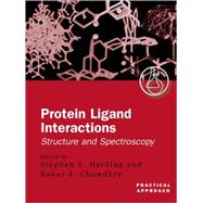
Protein-Ligand Interactions A Practical Approach Volume 2: Structure and Spectroscopy

by ;
  • ISBN13:

    9780199637478

  • ISBN10:

    0199637474

  • Edition: 1st
  • Format: Paperback
  • Copyright: 2001-03-22
  • Publisher: Oxford University Press
  • Purchase Benefits
List Price: $144.00

Summary

The two Practical Approach volumes on protein-ligand interaction do not comprise a comprehensive compilation of all the methods that can be used to investigate protein-ligand interactions. Instead, they are a selection of the most useful and easily applied methods and will be an invaluableguide to the principal techniques used to study the interactions of proteins and ligands. This second volume covers the major spectroscopic methods: FTIR, Raman, and fluorescence spectroscopy; circular dichroism, NMR, mass spectrometry, atomic force microscopy, and the use of paramagnetic probes.There are also chapters on X-ray crystallography and molecular modelling. Hydrodynamic and calorimetric techniques are covered in volume one. Both volumes are available individually, or as a set. Both volumes are written from a practical standpoint to be applicable to both academic and industrialscientists wishing to characterize protein-ligand systems by using a multi-disciplinary approach.

Table of Contents

List of protocols
xvii
Abbreviations xxi
X-ray crystallographic studies of protein-ligand interactions
1(98)
Rex A. Palmer
Introduction
1(1)
Crystallization of proteins and complexes for X-ray analysis
1(18)
Introduction
1(3)
Crystallization of proteins
4(1)
Practical considerations
5(10)
Crystal mounting for X-ray data collection
15(1)
Trouble shooting
16(1)
Protein-ligand complex crystals
17(2)
Crystal geometry and symmetry
19(10)
The crystal lattice
19(8)
Symmetry elements, crystal systems, and space groups
27(2)
X-ray diffraction from crystals
29(8)
Bragg's treatment of X-ray diffraction: the location of diffraction maxima
29(2)
The reciprocal lattice concept defining the location of diffraction maxima
31(2)
The structure factor F(hkl): X-ray intensities I(hkl)
33(3)
Calculated structure factors and R-factors
36(1)
Recording and measurement of X-ray diffraction patterns
37(9)
X-ray sources
37(1)
Recording and measuring intensity data
38(5)
Image plate data processing
43(3)
Preliminary crystal data
46(6)
Determination of unit cell and space group
46(2)
Resolution
48(1)
Number of reflections in the data set
48(1)
Completeness of a data set at a given resolution
48(1)
Internal consistency
49(1)
Number of molecules per unit cell
49(3)
Electron density: reconstruction of the molecular structure
52(3)
Basic principles
52(1)
Properties of the electron density
52(1)
Complementarity of the structure factor F(hkl) and electron density p(x, y, x)
53(1)
Difference electron density functions
53(1)
Patterson's Fourier series: the Patterson function P(uvw)
53(2)
Properties of the Patterson function
55(1)
Determination of the phases &phis;(hkl) for protein crystals
55(19)
Introduction
55(1)
Isomorphous replacement (IR)
56(1)
Heavy atoms and compounds for isomorphous replacement
56(1)
Preparation and screening of heavy atom derivatives
56(1)
Phase determination in single isomorphous replacement (SIR)
57(1)
Location of the heavy atoms
58(1)
Phase determination in multiple isomorphous replacement (MIR)
58(2)
Molecular replacement (MR)---the Sledgehammer
60(11)
Use of phase information and density modification
71(3)
What to do once phasing is acceptable
74(1)
Structure refinement, ligand fitting, and geometry verification
74(6)
Refinement
74(1)
Fourier refinement and simulated annealing
75(2)
Least squares refinement
77(3)
Examples of protein-ligand complex studies
80(13)
N-acetyl-DL-homocysteinyl ribonuclease derivatives: protein-ligand covalent modification
80(1)
Environment of the SO42- ion and solvent waters in native bovine RNase A active site at 0.9 A resolution (18)
81(1)
Enzyme-inhibitor interactions (non-covalent): r-rat RNase A binding with 2'-5-CpG (85)
82(1)
Protein-carbohydrate interactions: the non-covalent sugar binding sites in the lectin MLI
83(3)
Glycoproteins: covalently bound sugars and the [... Asn, X, Ser/Thr...] binding site; MLI B-chain Asn 136 sugar binding site (89)
86(1)
Protein-steroid interactions: Fab' fragment of antibody DB3 bound to progesterone and progesterone-like steroids
86(5)
Protein-nucleic acid interactions: procaryotic recognition of helix-turn-helix motifs in DNA (117)
91(2)
Further recommended reading and software
93(6)
Protein-peptide interactions: cyclophilin/cyclosporin complexes
93(1)
Protein-protein interactions (e.g. protein-antibody): camel VH with lysozyme
93(1)
Recommended books
93(1)
International tables
93(1)
Synchrotrons Web Page
93(1)
Space group diagrams
93(1)
Software for data processing
94(1)
Software for Fourier and structure factor calculations
94(1)
Software for MIR
94(1)
Software for packing and molecular geometry
94(1)
Software for graphics and model building
94(1)
Software for molecular graphics and display
94(1)
Software for refinement
94(1)
Software for molecular dynamics and minimization
94(1)
Databases
94(1)
References
95(4)
Molecular modelling
99(24)
Romano T. Kroemer
Introduction
99(1)
Protein structure prediction
100(4)
Ab initio predictions
101(1)
Threading, or fold recognition techniques
101(1)
Homology (or comparative) modelling
101(3)
Analysis of protein structures
104(1)
Docking of ligands to proteins
105(4)
Ab initio (de novo) ligand design
109(5)
Active site preparation
110(1)
Generation of structures
111(1)
Evaluation of structures
112(2)
Calculation of binding energies
114(2)
Statistical analysis of a set of protein ligands
116(1)
Concluding remarks
117(6)
Acknowledgements
117(1)
References
117(6)
Circular dichroism
123(46)
Alex F. Drake
Introduction
123(3)
The ordinary absorption of light
126(2)
The spectroscopic basis of circular dichroism
128(4)
The circular dichroism spectrometer
132(4)
The merits and uses of CD spectroscopy
136(2)
Protein/peptide conformations as studied by CD spectroscopy
138(7)
Instrumental measurement factors
143(1)
Spectroscopic factors
143(1)
Data analysis methods
144(1)
Monitoring ligand binding by CD spectroscopy
145(7)
Measurement conditions and instrumental parameters
146(1)
Further aspects of the concentration used for absorption and CD measurements
147(5)
Analysis of CD ligand binding titration studies
152(12)
Case IA
153(2)
Case IA'
155(2)
Case IB
157(1)
Case IB'
157(3)
The effect of concentration on binding constant determination
160(4)
CD kinetics---drug binding to proteins
164(5)
References
166(3)
Generic techniques for fluorescence measurements of protein-ligand interactions; real time kinetics and spatial imaging
169(32)
Josep Cladera
Paul O'Shea
Introduction
169(2)
Ligand-protein interactions revealed by intrinsic (tryptophan) fluorescence
171(1)
Tryptophan fluorescence measurements to demonstrate ligand-protein interactions
172(3)
Steady state tryptophan fluorescence quenching
172(1)
Time-resolved fluorescence lifetime and anisotropy decay measurements of albumin
172(3)
The surface electrostatic potential: one of the trinity of electrical membrane potentials
175(1)
Membrane electrostatics: implementation of fluoresceinphosphatidylethanolamine (FPE) to measure ligand-receptor interactions of membranes
175(4)
Labelling human cells with FPE
178(1)
Location of FPE in the membrane
178(1)
Digital imaging: using CCD fluorescence microscopy and confocal scanning laser fluorescence microscopy (CSLM) of ligand binding reactins of living cells
179(1)
Fluorescent images corrected for probe disposition and photobleaching
180(2)
Spatial imaging of the cell surface to identify localized ligand binding
182(1)
Experimental protocols
182(7)
Case studies
189(9)
Ligand (oleate) binding to HAS revealed by tryptophan fluorescenece quenching
189(1)
Kinetics of oleate-dependent fluorescence changes of HSA
190(1)
Interactions of a signal peptide (p25) with model membranes
190(3)
The interactions of serum albumin with B and T lymphocytes
193(1)
Receptor-mediated membrane binding of albumin
193(2)
Influence of interleukin-8 (IL-8) on the interaction of the gp41 fusion domain of HIV with T lymphocytes
195(1)
The interaction of Alzheimer's peptides with B12 microgial cells
196(2)
Quantitation of spatially localized interactions of ligands (albumin) with living cells
198(1)
Conclusions
198(3)
Acknowledgements
199(1)
References
199(2)
Stopped-flow techniques
201(38)
John F. Eccleston
Jon P. Hutchinson
Howard D. White
Introduction
201(1)
First- and second-order reactions
202(4)
First-order irreversible reactions
202(1)
Second-order irreversible reaction
203(1)
First-order reversible reaction
204(1)
Second-order reversible reactions
204(2)
Principle of operation of the stopped-flow instrument
206(2)
Testing the performance of a stopped-flow instrument
208(4)
The mixing process
208(1)
Mixing efficiency
208(2)
Measurement of dead-time
210(2)
Optimization of instrument
212(4)
Lamp selection
212(1)
Monochromator
212(1)
Choice of emission filter
213(1)
Stop volume
214(1)
Time constant
215(1)
Probes to monitor stopped-flow reactions
216(2)
Intrinsic probes
216(1)
Extrinsic probes
217(1)
Measurement of association and dissociation rate constants
218(4)
Initial consideration
218(1)
Association reactions
219(1)
Displacement reactions
219(1)
Example of determination of association and dissociation rate constants
220(1)
Kinetics of interaction of a non-fluorescent ligand with a protein
220(2)
Complex binding reactions
222(1)
Specialized techniques
222(9)
Double mixing
223(1)
Variable volume mixing
223(1)
Logarithmic time base
224(1)
Diode array detection
224(2)
Fluorescence anisotropy
226(5)
Modern continuous flow techniques
231(1)
Global analysis
231(8)
Strengths and limitations
232(1)
Example: myosin-subfragment 1.ADP binding to pyrene-actin
232(4)
Acknowledgements
236(1)
References
237(2)
Protein-ligand interactions studied by FTIR spectroscopy: methodological aspects
239(26)
Michael Jackson
Henry H. Mantsch
Introduction
239(1)
Protein structure determination by FTIR spectroscopy
240(2)
Experimental design
242(14)
Experimental technique
242(5)
Experimental conditions: choice of solvent
247(3)
Solvent and water vapour subtraction
250(3)
Derivation and deconvolution
253(3)
Thermal and solvent manipulation techniques for assessing protein-ligand interactions
256(4)
Difference spectroscopy
260(1)
Isotope edited difference spectroscopy
260(1)
Reaction-induced difference spectroscopy
261(4)
References
262(3)
Protein-ligand interactions studied by Raman and resonance Raman spectroscopy
265(46)
Robert Withnall
Introduction
265(3)
Raman experiments
268(24)
Visible/near infrared Raman spectroscopy
268(7)
FT Raman spectroscopy
275(2)
Difference Raman spectroscopy
277(2)
Resonance Raman spectroscopy
279(6)
Ultra-violet Raman and ultra-violet resonance Raman (UVRR) spectroscopy
285(1)
Raman optical activity (ROA)
286(2)
Time-resolved resonance Raman (TR3) spectroscopy
288(1)
Surface-enhanced Raman spectroscopy (SERS) and surface-enhanced resonance Raman spectroscopy (SERRS)
289(2)
Coherent anti-Stokes Raman spectroscopy (CARS)
291(1)
Interpretation of protein spectra and band assignments
292(3)
Amide group frequencies
292(3)
Group frequencies of side groups
295(1)
Specific information on proteins and their interactions with ligands
295(16)
Secondary structure
295(3)
Solvent exposure of aromatic side chains
298(1)
pKD's of protein-ligand complexes
299(1)
pKa's of specific protein groups
299(1)
Entropic and enthalpic changes on ligand binding
300(2)
Binding rate constants
302(4)
Acknowledgements
306(1)
References
307(4)
Electrospray ionization mass spectrometry
311(36)
T. J. Hill
D. Lafitte
P. J. Derrick
Introduction
311(1)
Mass spectrometry
311(2)
Electrospray ionization
313(14)
Principle
313(4)
Sample preparation
317(1)
Sample injection
318(2)
Creating a vacuum
320(1)
Mass analysis---selection and detection
320(3)
Data analysis
323(4)
Data storage
327(1)
Protein-ligand interactions of calmodulin
327(20)
Preparation of calmodulin
327(1)
ESI mass spectrometry of calmodulin
328(16)
References
344(3)
Nitroxide spin labels as paramagnetic probes
347(36)
David A. Middleton
Introduction
347(1)
Origin of the EPR spectrum
348(10)
Magnetic resonance
348(1)
Instrumentation
349(1)
Phase-sensitive detection and the first derivative spectrum
350(1)
Obtaining a spectrum
351(2)
EPR spectroscopy of nitroxide spin labels
353(5)
Conventional EPR spectroscopy
358(12)
Sample requirements
358(2)
Modification of protein side groups
360(8)
Analysis of multiple component spectra
368(2)
Saturation transfer EPR
370(7)
Aims
370(1)
Theory
371(1)
Recording a spectrum
372(3)
Interpretation of ST-EPR spectra
375(2)
Protein-ligand interactions
377(6)
Experimental design
377(3)
Time-resolved spectroscopy
380(1)
References
380(3)
NMR studies of protein-ligand interactions
383(24)
Lu-Yun Lian
Introduction
383(1)
Preparation of sample and preliminary binding studies
384(1)
Chemical exchange and analysing the NMR spectra for protein-ligand complexes
385(3)
Screening for ligand interactions
388(2)
Overview of NMR techniques used to study protein-ligand interactions
390(2)
Exchange rate measurements
392(3)
Assignment of resonances
395(2)
Protein resonance assignments
395(1)
Ligand resonance assignments
396(1)
Structural information
397(10)
Chemical shift changes
398(2)
Nuclear Overhauser effects and structure calculations
400(1)
Determination of ionization states
401(1)
Protein ionization states
402(3)
References
405(2)
Quantification and mapping of protein-ligand interactions at the single molecule level by atomic force microscopy
407(18)
Clive J. Roberts
Stephanie Allen
Martyn C. Davies
Saul J. B. Tendler
Philip M. Williams
Introduction
407(3)
Applications
410(1)
Quantification of the adhesion forces of individual protein-ligand complexes
410(11)
General guidelines
410(2)
Measurement of antibody-antigen interactions
412(9)
Spatial mapping of protein-ligand interactions
421(1)
Conclusions
421(4)
References
422(3)
List of suppliers 425(8)
Index 433

Supplemental Materials

What is included with this book?

The New copy of this book will include any supplemental materials advertised. Please check the title of the book to determine if it should include any access cards, study guides, lab manuals, CDs, etc.

The Used, Rental and eBook copies of this book are not guaranteed to include any supplemental materials. Typically, only the book itself is included. This is true even if the title states it includes any access cards, study guides, lab manuals, CDs, etc.

Rewards Program