did-you-know? rent-now

Amazon no longer offers textbook rentals. We do!

did-you-know? rent-now

Amazon no longer offers textbook rentals. We do!

We're the #1 textbook rental company. Let us show you why.

9780125441636

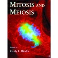
Methods in Cell Biology: Mitosis and Meiosis

by ; ;
  • ISBN13:

    9780125441636

  • ISBN10:

    0125441630

  • Format: Hardcover
  • Copyright: 1999-01-01
  • Publisher: Elsevier Science Serials
  • Purchase Benefits
  • Free Shipping Icon Free Shipping On Orders Over $35!
    Your order must be $35 or more to qualify for free economy shipping. Bulk sales, PO's, Marketplace items, eBooks and apparel do not qualify for this offer.
  • eCampus.com Logo Get Rewarded for Ordering Your Textbooks! Enroll Now
List Price: $180.00
  • Digital
    $196.88
    Add to Cart

    DURATION
    PRICE

Supplemental Materials

What is included with this book?

Summary

Mitosis and Meiosis details the wide variety of methods currently used to study how cells divide as yeast and insect spermatocytes, higher plants, and sea urchin zygotes. With chapters covering micromanipulation of chromosomes and making, expressing, and imaging GFP-fusion proteins, this volume contains state-of-the-art "how to" secrets that allow researchers to obtain novel information on the biology of centrosomes and kinetochores and how these organelles interact to form the spindle. Chapters Contain Information On: * How to generate, screen, and study mutants of mitosis in yeast, fungi, and flies * Techniques to best image fluorescent and nonfluorescent tagged dividing cells * The use and action of mitoclastic drugs * How to generate antibodies to mitotic components and inject them into cells * Methods that can also be used to obtain information on cellular processes in nondividing cells

Table of Contents

Contributors xi(4)
Preface xv
1. Isolation of Centrosomes from Drosophila Embryos
1(13)
Michelle Moritz
Bruce M. Alberts
I. Introduction
1(1)
II. Isolation of Centrosomes from Drosophila Embryos
2(3)
III. Assays for Microtubule Nucleation by Isolated Centrosomes
5(6)
IV. Conclusions
11(1)
References
11(3)
2. Studying the Composition and Function of Centrosomes in Vertebrates
14(22)
Michel Bomens
Mohammed Moudjou
I. Introduction
14(1)
II. Isolation of Centrosomes from Animal Cells
15(3)
III. Preparation of Immunological Probes from Isolated Centrosomes
18(2)
IV. Ultrastructural Analysis of Isolated Centrosomes
20(4)
V. Biochemical Composition of Centrosomes
24(3)
VI. Functional Assays of Isolated Centrosomes
27(2)
VII. Prospects
29(2)
References
31(5)
3. Isolation of Centrosomes from Spisula solidissima Oocytes
36(21)
Robert E. Palazzo
Jacalyn M. Vogel
I. Introduction
36(1)
II. Obtaining Organisms
37(1)
III. Isolation and Activation of Spisula solidissima Oocytes
37(4)
IV. Preparation of Oocyte Lysates
41(2)
V. Preparation of Microtubule Protein
43(2)
VI. Isolation of Centrosomes from Activated Oocyte Lysates
45(4)
VII. Immunofluorescence of Centrosomes and Asters
49(3)
VIII. Electron Microscopy of Asters and Centrosomes
52(1)
IX. Summary
53(3)
References
56(1)
4. Methods for in Situ Localization of Proteins and DNA in the Centromere-Kinetochore Complex
57(25)
A. Van Hooser
B.R. Brinkley
I. Introduction
57(2)
II. In Situ Localization of Proteins: Indirect Immunofluorescence
59(4)
III. In Situ Localization of Proteins: Immunogold EM
63(3)
IV. Fluorescent in Situ Hybridization Using DNA Satellite Probes
66(3)
V. Combination Staining: DNA/Protein
69(3)
VI. Specialized Techniques
72(6)
References
78(4)
5. Three-Dimensional Transmission Electron Microscopy and Its Application to Mitosis Research
82(32)
Bruce F. McEwen
Michael Marko
I. Introduction
82(2)
II. Resolution and Choosing between Tomography and Serial Sections
84(3)
III. Electron Tomography
87(11)
IV. Serial Section Reconstruction
98(1)
V. Analysis and Display of 3D Reconstructions
99(6)
VI. Software Packages
105(1)
VII. Summary and Conclusions
106(2)
References
108(6)
6. Enlightening Mitosis: Construction and Expression of Green Fluorescent Protein Fusion Proteins
114(23)
Kevin F. Sullivan
I. Introduction: Visualizing the Molecular Anatomy of the Spindle
114(1)
II. Fluorescence Properties of GFP
114(4)
III. Strategies for Constructing Fusion Proteins
118(5)
IV. Expression in Mammalian Cells
123(10)
References
133(4)
7. Recombinant p50/Dynamitin as a Tool to Examine the Role of Dynactin in Intracellular Processes
137(8)
Torsten Wittmann
Tony Hyman
I. Introduction
137(1)
II. Production of Recombinant p50/Dynamitin
138(2)
III. Disruption of the Dynactin Complex by p50/Dynamitin in Xenopus Egg Extracts
140(2)
IV. Disruption of Spindle Poles Using p50/Dynamitin
142(1)
References
142(3)
8. In Vitro Assays for Studying Saccharomyces cerevisiae Kinetochore Activity
145(11)
Fedor Severin
Ken Kaplan
Peter Sorger
Tony Hyman
I. Introduction
145(1)
II. Microtubule-Binding Assays for S. cerevisiae Kinetochores
146(5)
III. Band Shift Assay for the Kinetochore Complex
151(2)
References
153(3)
9. Fluorescent Speckle Microscopy of Spindle Microtubule Assembly and Motility in Living Cells
156(19)
Clare Waterman-Storer
Arshad Desai
E.D. Salmon
I. Introduction
156(1)
II. Principles of the Fluorescence Speckle Method for Microtubules
156(2)
III. Specimen Methods
158(3)
IV. Microscopy and Image Acquisition
161(3)
V. Image Processing and Analysis
164(3)
VI. Examples
167(3)
VII. Future Considerations
170(2)
References
172(3)
10. Polarized Light Microscopy of Spindles
175(34)
Rudolf Oldenbourg
I. Introduction
175(3)
II. Polarized Light Microscopy
178(12)
III. Analysis of Spindle Birefringence
190(7)
IV. Optimum Cell Types for Polarized Light Microscopy of Spindles
197(9)
References
206(3)
11. Micromanipulation of Chromosomes and Spindles in Insect Spermatocytes
209(10)
Dahong Zhang
R. Bruce Nicklas
I. Introduction
209(1)
II. Preparing for Micromanipulation
210(6)
III. Manipulating Cell Components
216(1)
References
217(2)
12. Microinjection of Mitotic Cells
219(14)
Patricia Wadsworth
I. Introduction
219(2)
II. Choice of Cells
221(2)
III. Timing of Injection
223(1)
IV. Microinjection Procedure
223(5)
V. Conclusion
228(1)
References
229(4)
13. Obtaining Antibodies to Spindle Components
233(12)
Ryoko Kuriyama
Kathy Ensrud
I. Introduction
233(2)
II. Methods
235(8)
III. Discussion
243(1)
References
244(1)
14. Using Antisense Technology to Study Mitosis
245(22)
Linda Wordeman
Mike Wagenbach
I. Introduction
245(2)
II. Antisense Mechanism of Action
247(1)
III. Choice of Antisense Reagents
248(6)
IV. Assaying Target Protein Levels
254(1)
V. Antisense Reagents Used to Study Cell Division
255(6)
References
261(6)
15. The Use and Action of Drugs in Analyzing Mitosis
267(30)
Mary Ann Jordan
Leslie Wilson
I. Introduction: Why Use Drugs?
267(1)
II. Brief Overview of Microtubule Assembly Dynamics
268(2)
III. Mechanisms of Action of Major Antimitotic Drugs: Binding to Tubulin and Microtubules and Effects on Microtubule Polymerization and Dynamics and on Mitosis
270(17)
IV. Determination of Intracellular Drug Levels
287(1)
V. How to Use Antimitotic Drugs: Practical Guidelines
288(1)
References
289(8)
16. Correlative Light and Electron Microscopy of Mitotic Cells in Monolayer Cultures
297(21)
Conly L. Rieder
Grisel Cassels
I. Introduction
297(2)
II. Light Microscopy
299(3)
III. Flat Embedding
302(3)
IV. Preparing the Cell for Sectioning
305(2)
V. Obtaining the Required Ultrastructural Information
307(6)
References
313(5)
17. Identification and Characterization of Mitotic Mutations in Drosophila
318(30)
William E. Theurkauf
Margarete M. S. Heck
I. Introduction
318(4)
II. Maternal-Effect Mutations That Disrupt the Syncytial Mitotic Divisions
322(2)
III. Cytological Analysis of the Syncytial Mitoses
324(10)
IV. Zygotic Mutations That Disrupt Mitosis in Larval Tissues
334(3)
V. Cytological Analysis of Larval Brain and Imaginal Discs
337(7)
References
344(4)
18. Methods for Isolating and Analyzing Mitotic Mutants in Aspergillus nidulans
348(21)
Berl R. Oakley
I. Introduction
348(1)
II. Characteristics of Aspergillus nidulans
348(5)
III. Strains and Media
353(1)
IV. Harvesting Conidia and Preparing Conidial Suspensions
354(1)
V. Mutagenesis
355(1)
VI. Methods for Isolating Mitotic Mutants in A. nidulans
355(6)
VII. Morphological Analysis of Mutants
361(2)
VIII. Genetic Analysis of Mutants
363(2)
IX. Molecular Genetic Methods for Working with A. nidulans
365(2)
References
367(2)
19. Using Green Fluorescent Protein Fusion Proteins to Quantitate Microtubule and Spindle Dynamics in Budding Yeast
369(17)
Kerry Bloom
Dale L. Beach
Paul Maddox
Sidney L. Shaw
Elaine Yeh
E. D. Salmon
I. Introduction
369(1)
II. Construction of Protein-GFP Fusion and Promoter Selection
370(3)
III. Quantifying Fluorescence in Cell Populations
373(2)
IV. The Imaging System
375(1)
V. Quantitative Solution to the Imaging Problem
376(2)
VI. Image Acquisition and Processing
378(1)
VII. Applications and Examples: Expression of Dynein-GFP in Vivo
379(2)
References
381(5)
20. The Use of Xenopus Egg Extracts to Study Mitotic Spindle Assembly and Function in Vitro
386(27)
Arshad Desai
Andrew Murray
Timothy J. Mitchison
Claire E. Walczak
I. Introduction
386(1)
II. Preparation of CSF Extracts for Spindle Assembly
387(6)
III. Spindle Assembly Reaction
393(4)
IV. Monitoring Spindle Assembly Reactions
397(4)
V. Manipulation of Extracts
401(3)
VI. Data Analysis and Interpretation
404(1)
VII. Anaphase in Vitro
405(5)
VIII. Conclusions
410(1)
References
411(2)
21. Methods for Studying Cell Division in Higher Plants
413(27)
Jan W. Vos
Aline H. Valster
Peter K. Hepler
I. Introduction
413(1)
II. Cell Types of the Study of Cell Division
414(14)
III. Microinjection of Plant Cells
428(5)
IV. Conclusions
433(1)
References
434(6)
22. Using Sea Urchin Gametes for the Study of Mitosis
440(33)
Greenfield Sluder
Fredrick J. Miller
Edward H. Hinchcliffe
I. Introduction
440(1)
II. The Experimental System
440(1)
III. Maintenance of the Organisms
441(2)
IV. Obtaining Gametes
443(1)
V. Zygotes
444(4)
VI. Mounting Cells for Observation
448(14)
VII. Other Methods
462(2)
VIII. Annotated List of References
464(8)
References
472(1)
Index 473

Supplemental Materials

What is included with this book?

The New copy of this book will include any supplemental materials advertised. Please check the title of the book to determine if it should include any access cards, study guides, lab manuals, CDs, etc.

The Used, Rental and eBook copies of this book are not guaranteed to include any supplemental materials. Typically, only the book itself is included. This is true even if the title states it includes any access cards, study guides, lab manuals, CDs, etc.

Rewards Program