Note: Supplemental materials are not guaranteed with Rental or Used book purchases.
Purchase Benefits
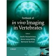
Looking to rent a book? Rent Textbook of in vivo Imaging in Vertebrates [ISBN: 9780470015285] for the semester, quarter, and short term or search our site for other textbooks by Ntziachristos, Vasilis; Leroy-Willig, Anne; Tavitian, Bertrand. Renting a textbook can save you up to 90% from the cost of buying.
Anne Leroy-Willig. Université Paris-Sud, France
Bertrand Tavitian. Unité d'Imagerie de l'Expression des Genes, CEA - SHFJ - INSERM ERITM, Orsay Cedex, France
| Contributors | |
| Introduction | |
| Nuclear Magnetic Resonance Imaging and Spectroscopy | |
| Introduction | |
| Magnets and magnetic field | |
| Nuclear magnetization | |
| Excitation and return to equilibrium of nuclear magnetization | |
| The NMR hardware: RF coils and gradient coils (more technology) | |
| NMR spectroscopy: the chemical encoding | |
| How to build NMR images: the spatial encoding | |
| MRI and contrast | |
| Sensitivity, spatial resolution and temporal resolution | |
| Contrast agents for MRI | |
| Imaging of 'other' nuclei | |
| More parameters contributing to MRI contrast | |
| More about applications | |
| High Resolution X-ray Microtomography: Applications in Biomedical Research | |
| Introduction | |
| Principles of tomography | |
| Implementation | |
| Contribution of microtomography to biomedical imaging | |
| Ultrasound Imaging | |
| Principles of ultrasonic imaging and its adaptation to small laboratory animals | |
| Pulse-echo transmission | |
| Ultrasonic transducers | |
| From echoes to images | |
| Blood flow and tissue motion | |
| Non-linear and contrast imaging | |
| Discussion | |
| Introduction | |
| Radioactivity | |
| Interaction of gamma rays with matter | |
| Radiotracer imaging with gamma emitters | |
| Detection of positron emitters | |
| Image properties and analysis | |
| Radiochemistry of gamma-emitting radiotracers | |
| Radiochemistry of positron-emitting radiotracers | |
| Major radiotracers and imaging applications | |
| Optical Imaging and Tomography | |
| Introduction | |
| Light - tissue interactions | |
| Light propagation in tissues | |
| Reconstruction and inverse problem | |
| Fluorescence molecular tomography (FMT) | |
| Optical Microscopy in Small Animal Research | |
| Introduction | |
| Confocal laser scanning microscopy | |
| Multiphoton laser scanning microscopy | |
| Variants for In vivo imaging | |
| Surgical preparations | |
| Applications | |
| New Radiotracers, Reporter Probes and Contrast Agents | |
| Introduction | |
| New radiotracers | |
| Multimodal constructs for magnetic resonance imaging | |
| Fluorescence reporters for biomedical imaging | |
| New contrast agents for NMR | |
| Imaging techniques - reporter gene imaging agents | |
| Multi-Modality Imaging | |
| Introduction | |
| Concurrent imaging versus computer-assisted registration | |
| Combination of SPECT and CT | |
| FMT registration with MRI | |
| Brain Imaging | |
| Introduction | |
| Bringing amyloid into focus with MRI microscopy | p. Annemie Van |
| Cerebral blood volume and BOLD contrast MRI unravels brain responses to ambient temperature fluctuations in fish | p. Annemie Van |
| Assessment of functional and neuroanatomical re-organization after experimental stroke using MRI | |
| Table of Contents provided by Publisher. All Rights Reserved. |
The New copy of this book will include any supplemental materials advertised. Please check the title of the book to determine if it should include any access cards, study guides, lab manuals, CDs, etc.
The Used, Rental and eBook copies of this book are not guaranteed to include any supplemental materials. Typically, only the book itself is included. This is true even if the title states it includes any access cards, study guides, lab manuals, CDs, etc.